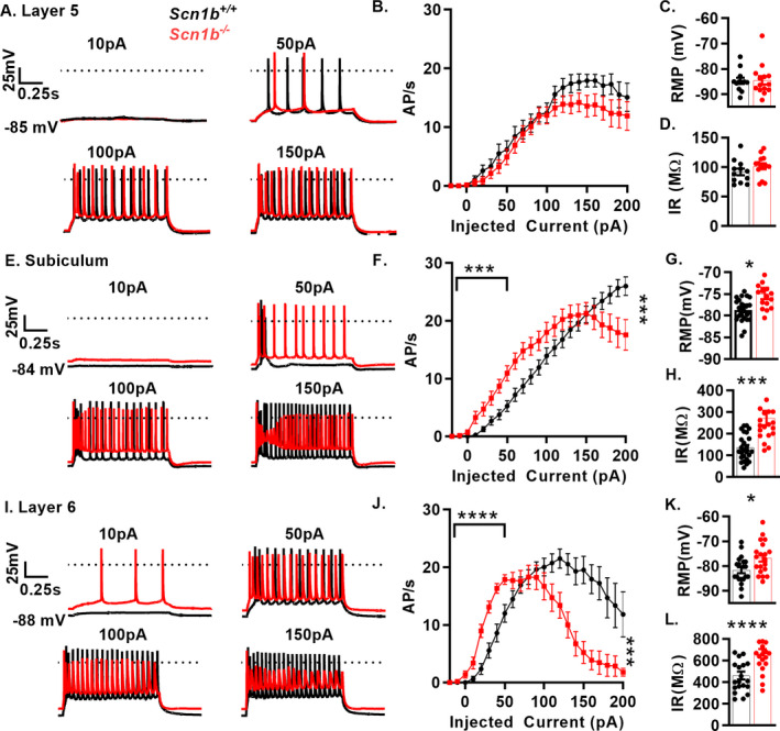
Figure 4.

Scn1b deletion results in complex pyramidal neuron excitability defects. A., E, and I. Representative voltage traces from whole‐cell recordings of layer 5 (A), subiculum (E) and layer 6 (I) pyramidal neurons in acute brain slices of Scn1b +/+ (black) or Scn1b ‐/‐ mice (red) B., F., J., Current injection vs. APs fired in 1‐s of recordings above. Firing at low current injection quantified as area under the curve up to 50 pA. Depolarization block is quantified as AP count at highest current injection divided by max AP count (with AP failure defined as max voltage < 0 mV, right asterisks). Layer 5 (B) pyramidal neurons show no change in firing at low current injections or degree of depolarization block (n/N = 12/7 Scn1b +/+, 14/7 Scn1b ‐/‐). Subicular (F) and Layer 6 pyramidal neurons (J) and show increased firing at low current amplitudes (left asterisks indicate p‐value) in Scn1b +/+ vs. Scn1b ‐/‐ mice (n/N = 25/12 Scn1b+/+, 24/13 Scn1b‐/‐ subiculum; n/N = 20/10 Scn1b +/+, 21/9 Scn1b ‐/‐ layer 6, Welch’s t‐test). C., G., K. RMP is not affected by Scn1b deletion in layer 5 (C) but is depolarized in subicular (G) and layer 6 pyramidal neurons (K). D., H., L. Input resistance is unaffected by Scn1b deletion in layer 5 (D) but is increased in subicular (H) and layer 6 pyramidal neurons (L). See Table 3 for quantification of biophysical properties. Asterisks indicate p‐values (*P < 0.05, ***P < 0.005, and ****P < 0.0001)
