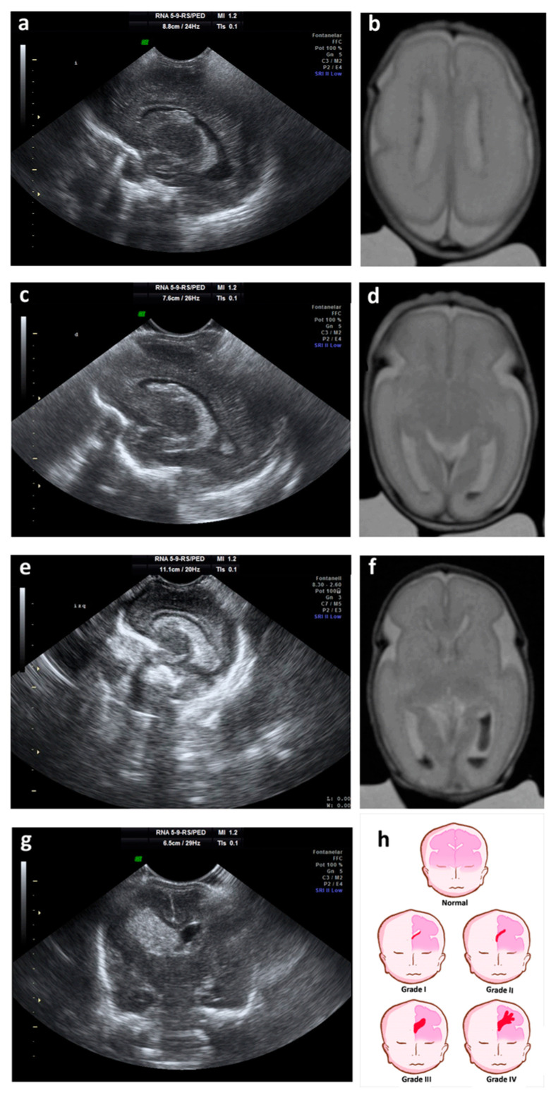
Figure 1.
Ultrasound and magnetic resonance imaging (MRI) showing different grades of GM-IVH. (a) Parasagittal cerebral ultrasound through lateral ventricles shows grade I hemorrhage; (b) T2-weighted axial MRI shows grade I hemorrhage on both lateral ventricles; (c) parasagittal cerebral ultrasound through lateral ventricles shows grade II hemorrhage; (d) T2-weighted axial MRI shows grade II hemorrhage on the left lateral ventricle; (e) parasagittal cerebral ultrasound through lateral ventricles shows grade III hemorrhage; (f) T2-weighted axial MRI shows grade III hemorrhage on the left lateral ventricle and grade II hemorrhage on the right lateral ventricles; (g) coronal cerebral ultrasound shows grade IV or periventricular hemorrhagic infarction; (h) cartoon representing ultrasound classification of the germinal matrix-intraventricular hemorrhage (GM-IVH).

