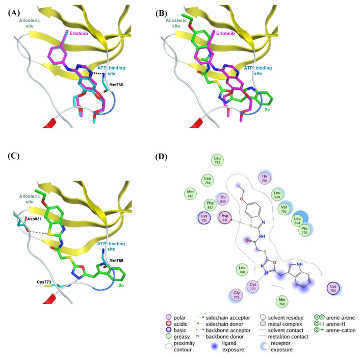
Figure 2.
The top-scoring docked pose of compound 2e to the EGFR active site (PDB code 4HJO) as predicted by MOE 2019.01. (A) Comparison of modeled binding mode of the co-crystallized ligand erlotinib (magenta sticks) and its superimposed docking conformation (cyan sticks). (B) Comparison of modeled binding mode of compound 2e (green sticks) and erlotinib (magenta sticks). (C) Detailed binding mode of compound 2e (green sticks) displaying hydrogen bonds (black dashed line) with the critical amino acid residues (cyan sticks). (D) Two-dimensional depiction of compound 2e binding interactions with the essential amino acid residues.

