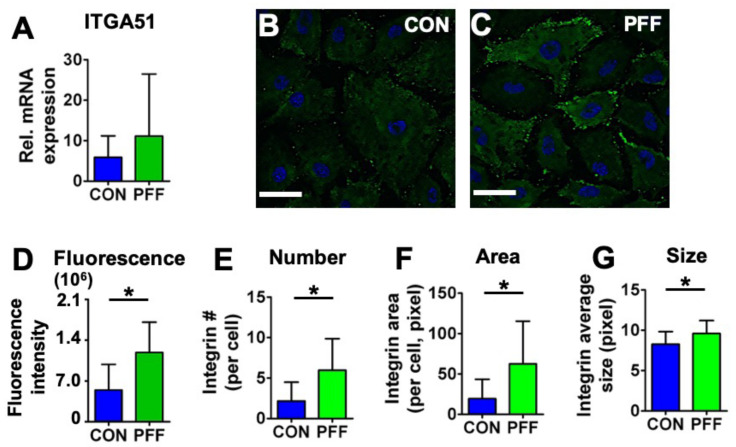
Figure 5.
Effect of 1 h PFF on integrin-α5 expression and distribution in MC3T3-E1 osteoblasts. (A) mRNA expression of ITGA51. (B) 2D images of control cells stained for integrin-α5 (green) and DAPI for the nucleus (blue). (C) 2D images of 1 h PFF-treated cells stained for integrin-α5 (green) and nucleus (blue). Bar = 50 µm. (D) Fluorescence intensity of integrin-α5 in control and PFF-treated cells. (E) Integrin-α5 number in control and PFF-treated cells. (F) Integrin-α5 area in control and PFF-treated cells. (G) The integrin-α5 average size in control and PFF-treated cells. * p < 0.05. CON, control; PFF, pulsating fluid flow.

