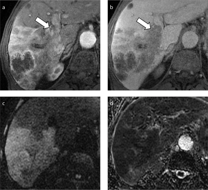
Figure 15. a–d.
An 82-year-old man with cirrhosis and history of HCC. MRI images on hepatic arterial (a) and portal venous (b) phases show a large HCC with infiltrative imaging appearance and macrovascular invasion (arrows) in the right portal vein. DWI image at b= 800 s/mm2 (c) demonstrates diffusion restriction of the liver mass and hypointensity on ADC map (d).

