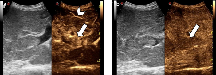
Figure 4. a, b.
An 84-year-old man with hepatitis B virus (HBV)-related cirrhosis and tumor thrombus. Contrast-enhanced ultrasound image (a) at 13 seconds after the intravenous administration of contrast agent demonstrates the presence of an enhancing thrombus within the main portal vein (arrow), simultaneously with the intra-hepatic artery (arrowhead). Image acquired at 180 seconds (b) shows washout of the tumor thrombus (arrow).

