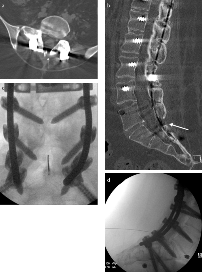
Figure 2. a–d.
A 21-year-old male SMA-3 patient with near complete fusion of posterior elements. Only a tiny nonfused area (white arrow) was present at L5-S1 on axial (a) and sagittal (b) CT reconstructions. This area could be visualized with fluoroscopy (c, d) when correlated with CT; however, patient’s sacral soft tissues were significantly thick, which required a 7-inch long needle. Initially, the first two injections lasted 7.2 and 4.6 min to complete. However, after the initial experience and familiarity with patient’s anatomy, the subsequent procedures were completed in less than 30 s.

