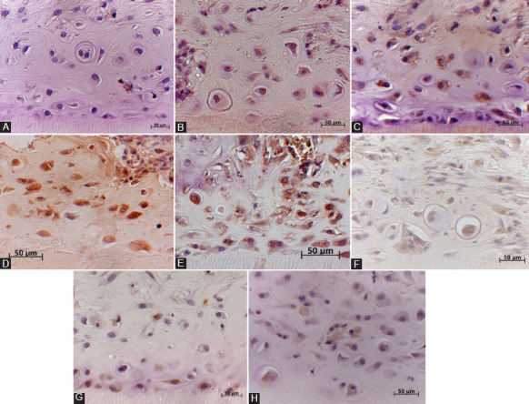
FIGURE 2.

(A) The cytoplasm of odontoblast/osteoblast-like cells formed by dental pulp cells (DPCs) in the collagen type 1 scaffold inside newly formed osteodentin-like hard tissue matrix near the dentinal wall and in the center of the dentin tubule, showing positive brown staining for alkaline phosphatase (ALP) at 21 days in the experimental group; (B) the cytoplasm of osteoblast-like cells inside the bone matrix formed by DPCs without a collagen gel shows positive staining for ALP in the control group at 21 days; (C) the cytoplasm of odontoblast/osteoblast-like cells inside the osteodentin-like matrix formed by DPCs in the collagen type 1 scaffold shows positive immunoreactivity for bone sialoprotein (BSP) at 21 days in the experimental group; (D) osteoblasts inside the bone matrix formed by DPCs show positive staining for BSP in the control group at 21 days; (E) the cytoplasm of odontoblast/osteoblast-like cells inside the osteodentin-like matrix formed by DPCs in the collagen type 1 scaffold shows positive immunoreactivity for osteopontin (OPN) at 21 days in the experimental group. OPN-positive osteoblast/odontoblast-like cells are observed throughout the dentin tubule and near the dentinal wall; (F) osteoblasts inside the bone matrix formed by DPCs show weak staining for OPN in the control group at 21 days; (G) the cytoplasm of odontoblast-like cells formed by DPCs in the collagen type 1 scaffold inside newly formed osteodentin-like hard tissue matrix shows positive brown staining for nestin at 21 days in the experimental group. Nestin-positive cells are observed near the dentinal wall of the demineralized dentin tubules; (H) osteoblast/odontoblast-like cells inside the bone matrix formed by DPCs show weak staining for nestin in the control group at 21 days.
