Figure 1.
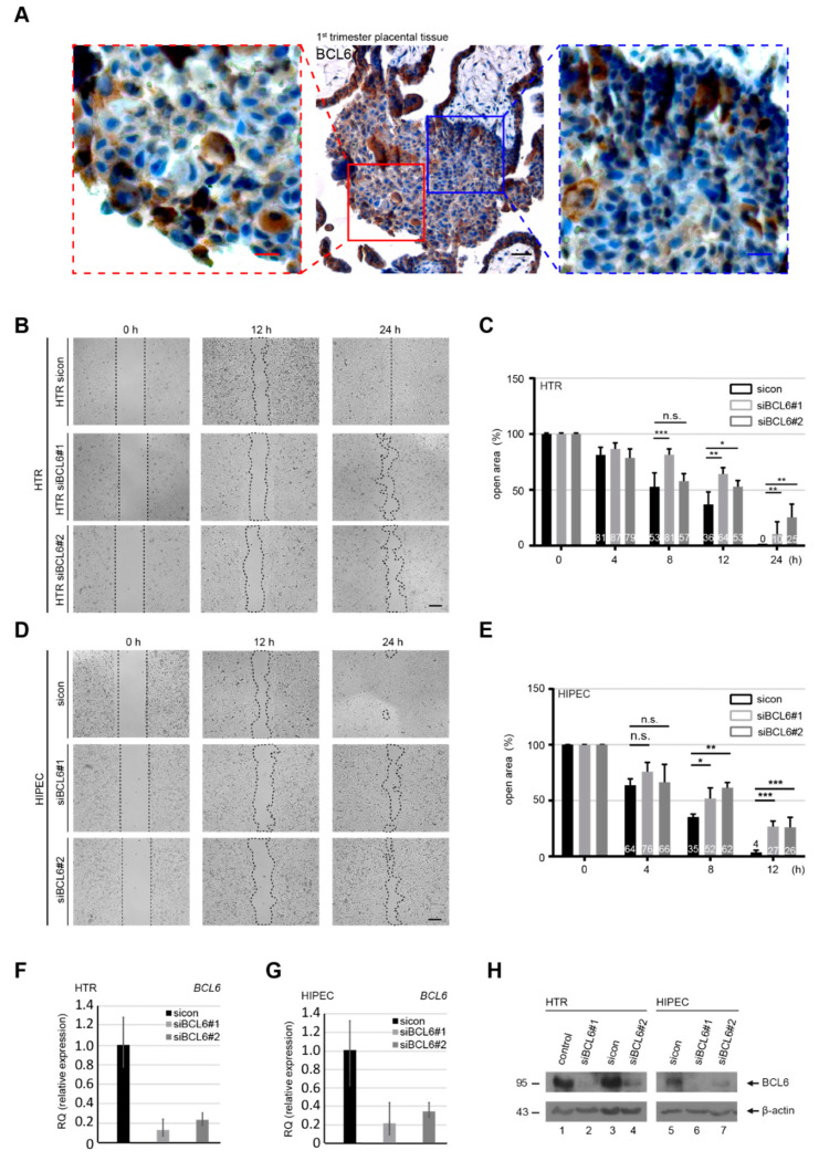
Abundance of B-cell lymphoma 6 (BCL6) in first trimester placentas and requirement for the migration of trophoblastic cells. (A) First trimester placental tissue sections were immunohistochemically stained for BCL6 and DNA. Representatives are shown. Scale: 50 μm, inset scale: 20 μm. (B,D) HTR-8/SVneo (HTR) (B) or HIPEC-65 (HIPEC) (D) cells were treated with control siRNA (sicon) or two different siRNAs targeting the coding region of BCL6 (siBCL6#1 and siBCL6#2) for 12 h. Treated cells were subjected to wound healing/migration assays. Migration front closure was imaged at 0, 8, and 12 h and bright-field images are shown. The migration front is visualized by black dashed lines. Scale: 300 μm. (C,E) The open areas of HTR (C) or HIPEC (E) cells were quantified and are presented in a bar graph at multiple time points. The reference point was the cell-free area at 0 h, which was set as 100%. Data were obtained from three independent experiments and are shown as the mean ± SEM. *** p < 0.001, ** p < 0.01, * p < 0.05 and, n.s. p > 0.05. (F–H) Relative BCL6 gene levels in treated HTR (F) and HIPEC (G) cells were measured as transfection efficiency controls, as well as the protein levels analyzed by Western blot.

