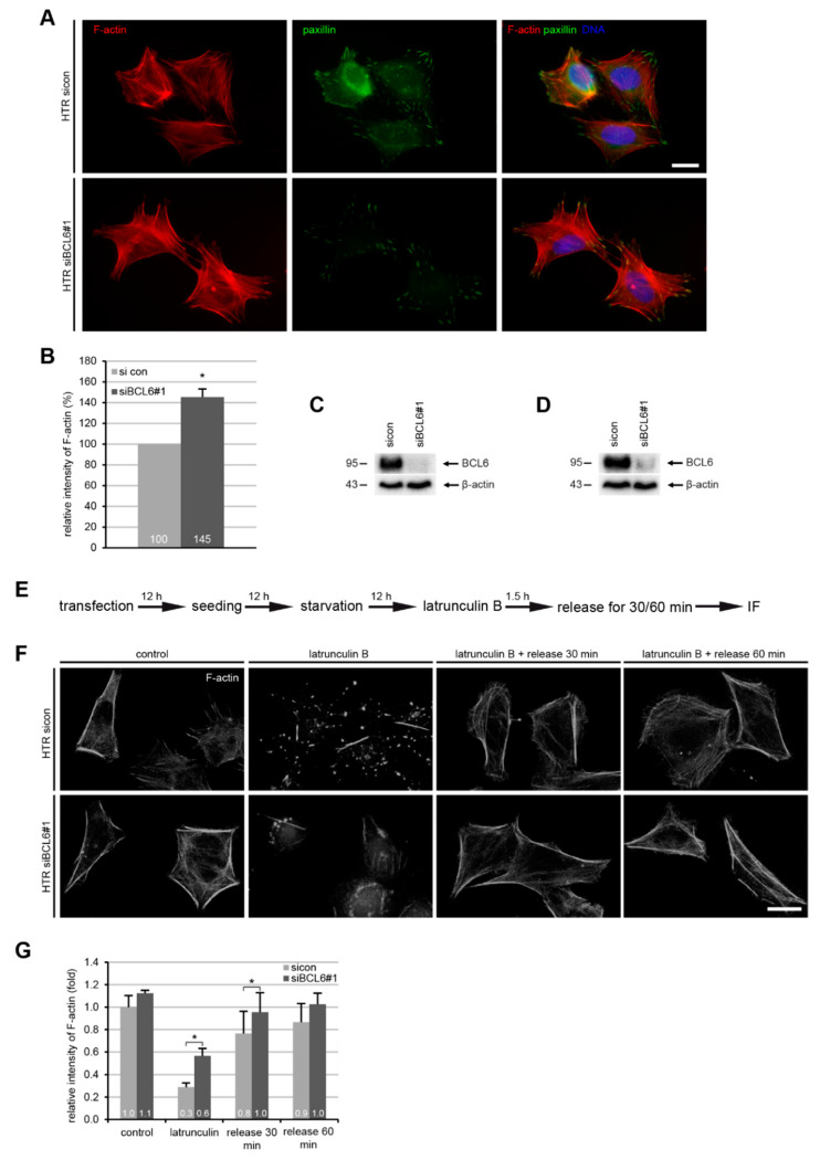
Figure 4.
Elevated F-actin and reduced paxillin in HTR cells with reduced BCL6. (A) HTR cells, treated with control siRNA (sicon) or siRNA targeting BCL6 (siBCL6#1) for 24 h, were stained for F-actin and paxillin for microscopic evaluation. Representatives are shown. Scale: 25 μm. (B) The relative intensity of F-actin was measured in treated HTR cells (200 cells for each condition). The results were obtained from three independent experiments and are presented as the mean ± SEM. * p < 0.05. (C) Western blot analysis as the transfection efficiency control for (A,B). Β-actin served as the loading control. (D) Western blot analysis as the transfection efficiency control for (F,G). β-actin served as the loading control. (E) Schedule of the latrunculin B washout experiment. HTR cells, transfected with sicon or siRNA BCL6#1 for 24 h, where 10 µM latrunculin B was added into the medium for 90 min, and after 30 and 60 min, cells were released into fresh prewarmed culture medium. (F) Cells were stained for F-actin (phalloidin, white). Representatives images of the reassembly rate of actin fibers are presented. Scale: 25 µm. (G) The mean fluorescence intensity of F-actin (phalloidin) was quantified and is presented (30 cells were used per condition). The experiment was independent repeated three times and the results are presented as the mean ± SEM. * p < 0.05.

