Figure 6.
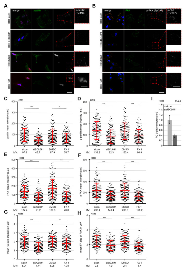
Reduced paxillin, p-paxillin, focal adhesion kinase (FAK), and phosphorylated FAK (p-FAK) at FAs of HTR cells deficient of BCL6. (A,B) HTR cells were treated with sicon or siBCL6#1, with DMSO or BCL6 inhibitor FX1. Twenty-four hours later, cells were stained for paxillin (green), p-paxillin (white), and DNA (blue, DAPI) (A), or for FAK (green), p-FAK (red), and DNA (blue, DAPI) (B). Representatives are shown. Scale: 25. Insets are magnified regions. Scale: 20. (C,D) Mean intensities of paxillin (C) and p-paxillin (D) were measured (280 FAs for each condition). The results were derived from three independent experiments and are presented as scatter plots with variations. a.u., arbitrary units. (E,F) Mean intensities of FAK (E) and p-FAK (F) were measured (280 FAs for each condition). The results were derived from three independent experiments and are presented as scatter plots with variations. a.u., arbitrary units. (G,H) Mean sizes of paxillin (G) or FAK (H) in FAs were measured (280 FAs for each condition). The results were obtained from three independent experiments and are presented as scatter plots with variations. (I) BCL6 gene analysis as the transfection efficiency control. For (C–H): *** p < 0.001, ** p < 0.01, and * p < 0.05.

