FIGURE 1.

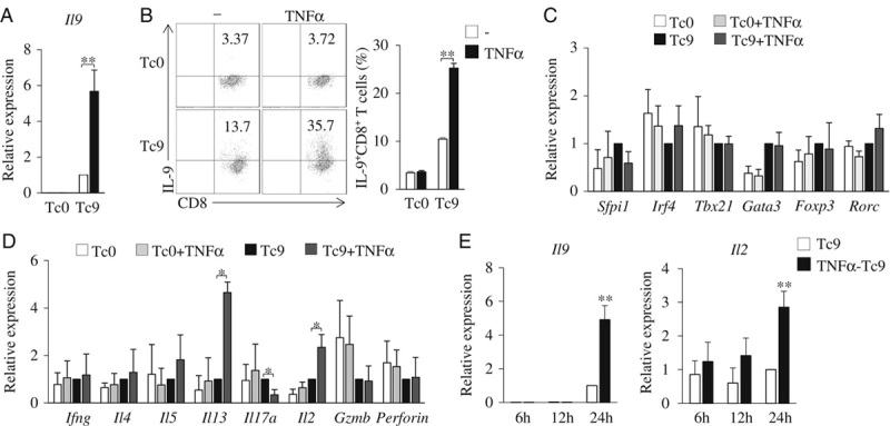
TNF-α promotes the induction of Tc9 cells. A–D, Mouse-naive CD8+ T cells were cultured under Tc9-polarizing conditions with or without the addition of TNF-α for 2 days. Cells cultured without the addition of TGF-β and IL-4 (Tc0) were used as controls. A, Quantitative polymerase chain reaction analysis of Il9 expression in CD8+ T cells. Expression was normalized to Gapdh and set at 1 in cells treated with TGF-β plus IL-4 (Tc9 cells). B, Flow cytometry analysis of IL-9-expressing CD8+ (IL-9+CD8+) T cells. Numbers in the dot plots represent the percentages of IL-9+CD8+ T cells. Right, summarized results of 3 independent experiments obtained as at left. Quantitative polymerase chain reaction analysis of the indicated transcription factors (C), cytokines, and effector molecules (D) in T cells. Expression was normalized to Gapdh and set at 1 in Tc9 cells. E, Mouse-naive CD8+ T cells were cultured under Tc9-polarizing conditions with or without the addition of TNF-α. Cells were collected at the indicated time points, and quantitative polymerase chain reaction analyzed the expression of Il9 and Il2 in Tc cells. Expression was normalized to Gapdh and set at 1 in Tc cells without the addition of TNF-α. Data are representative of 3 (B) independent experiments or presented as mean±SD of 3 (A–E) independent experiments. *P<0.05; **P<0.01. IL indicates interleukin; TGF, transforming growth factor; TNF, tumor necrosis factor.
