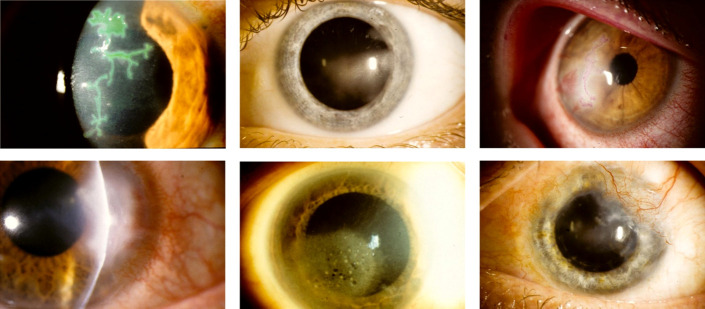
FIGURE 3.
Clinical photography of varying manifestations of HSV keratitis. Top row: epithelial keratitis with pathognomonic dendritic ulcer, visualized with lissamine green staining (left); stromal keratitis without ulceration (middle); mixed epithelial and stromal keratitis, visualized with rose bengal dye staining (right). Bottom row: stromal keratitis with ulceration (left); endothelial keratitis (middle); chronic, scarring stromal keratitis with limbal neovascularization (right).

