Figure 4.
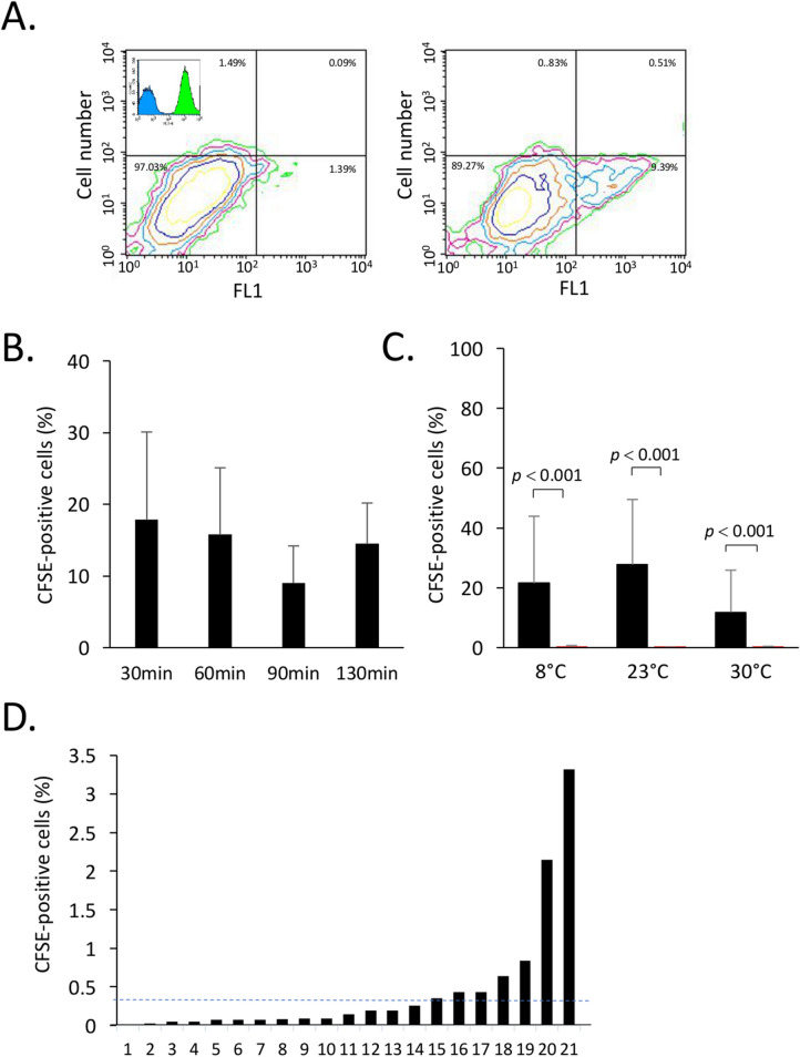
Entry of hemocytes in mussels. CFSE-stained hemocytes from Mytilus spp. were added to seawater. After the indicated times, IVF and hemolymph were collected and analyzed for the presence of CFSE-positive cells. (A) A representative contour plot showing hemocytes collected from IVF of mussels incubated without (left) and with CFSE-positive hemocytes (right). In the upper corner of the left contour plot, a representative overlay histogram of unstained (control) hemocytes in blue and CFSE stained hemocytes in green, (B) Effect of temperature on entry of CFSE-positive cells in IVF of mussels. (C) Percentage of CFSE-positive cells recovered in IVF of mussels at different temperatures 90 min after addition of CFSE-stained cells. The autofluorescence controls are shown in red. Its shows the autofluorescence as measured in (A) generated by unstained hemocytes recovered in IVF of mussels incubated in the same conditions. (D) Percentages of CFSE-positive cells present in hemolymph of Mytilus spp. maintained at 23 °C for 90 min after addition of CFSE-positive cells. The dotted line represents the average of CFSE-positive cells in hemolymph. The data are representative of two independent experiments.

