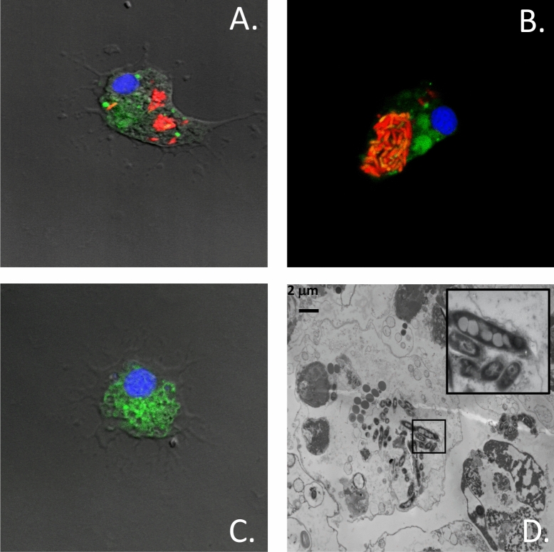
Figure 5.
Infection of hemocytes by M. marinum. (A,B) Hemocytes from Mytilus spp. were infected with mCherry-expressing M. marinum. Cells were incubated with the CFSE and H33342 prior to analysis by confocal microscopy. (C) Uninfected hemocytes (Control). (D) Transmission electron microscopy showing a representative hemocyte infected with M. marinum. The insert on the upper right shows an enlarged area of the infected cell. The data are representative of two independent experiments.

