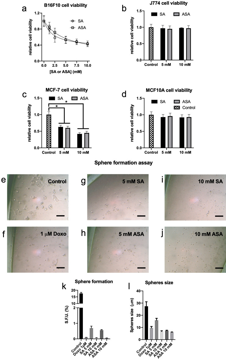
Figure 1.
SA and ASA reduced cancer cells viability and impeded sphere formation capability in a 3D culture model. B16F10 (Panel a), J774 (Panel b), MCF-7 (Panel c) and MCF10A (Panel d) cells were grown in 2D cultures and treated with the concentrations of SA or ASA indicated on the abscissa for 24 h. These results are presented as the mean ± S.E.M of 4 independent experiments (n = 4). Panels e–j: Representative optical microscopy pictures of 3D-cultured B16F10 cells untreated (b) or treated with 1 µM doxorubicin (c), 5 mM SA (d), 5 mM ASA (e), 10 mM SA (f) and 10 mM ASA (g). Panels k and l: quantification of the numbers and the size, respectively, of spheres formed. These results are represented as mean ± S.E.M of 3 independent experiments (n = 3). * means P < 0.05 as compared to the control (One-way ANOVA followed by Dunnett post-test).

