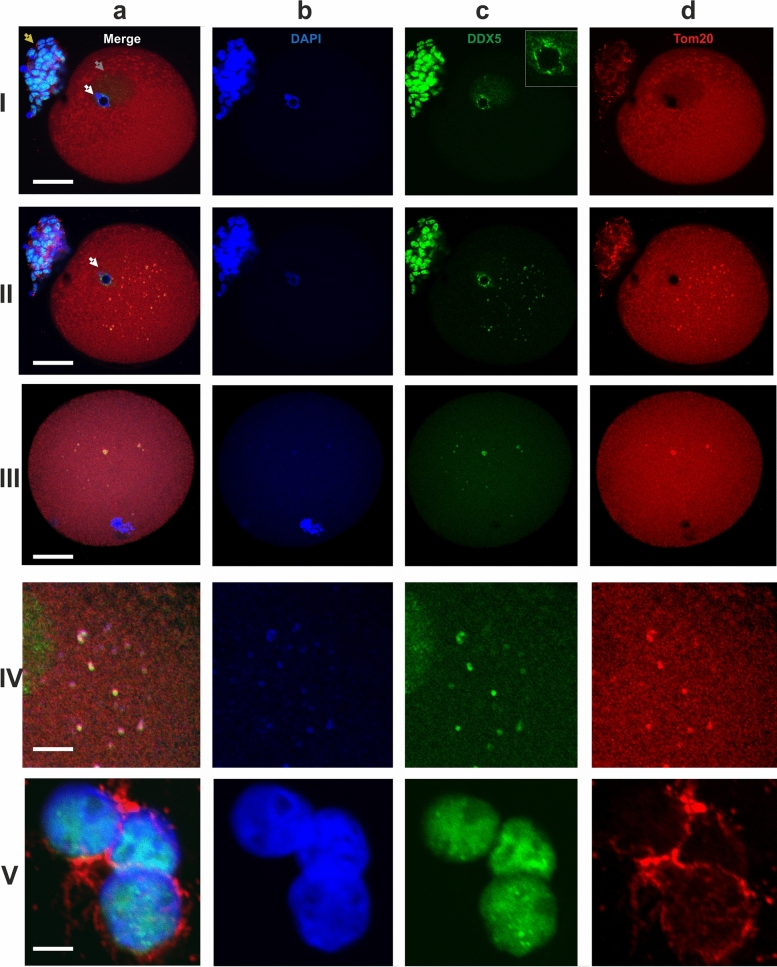
Figure 2.
Localization of the Tom20 (d, red) and DDX5 RNA helicase (c, green) in two different optical sections of a GV oocyte (I—intranuclear, II—ooplasmic DDX5), in an optical section of an MI oocyte (III) and in cumulus cells (V). The ooplasmic structures co-stained with the AB against Tom20 and DDX5 are shown at a higher magnification in (IV). The insert in the image 2-Ic shows a magnified area of heterochromatic ring stained with the antiDDX5 AB. White arrows—a ring of the highly condensed heterochromatin; gray arrow—the nucleus of a GV oocyte; yellow arrows—residual cumulus cells. Chromatin is counterstained with DAPI (b, blue). The pseudocolored merged images are shown in (a). Scale bars—50 μm (I–III), 10 μm (IV–V).

