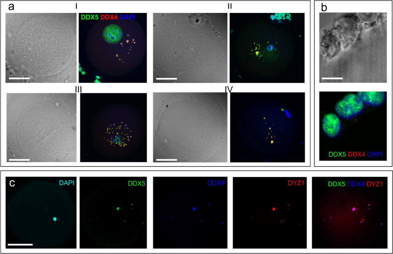
Figure 3.
Localization of the DDX5 (a–c, green) and DDX4 (a,b—red, c—blue) RNA helicases in GV (a, I), late GV (a, II and c), GVBD (a, III) and MI (a, IV) oocytes) and in cumulus cells (b). Chromatin is counterstained with DAPI (a,b, blue; c—cyan). Pseudocolored merged images of a single optical section are shown in (a), (b). Black and white images in (a), (b) represent phase contrast images taken simultaneously with the corresponding optical section. The overlapping of helicases with DYZ1 DNA–RNA FISH signals is shown in c. Scale bars—50 μm (a,c), 10 μm (b).

