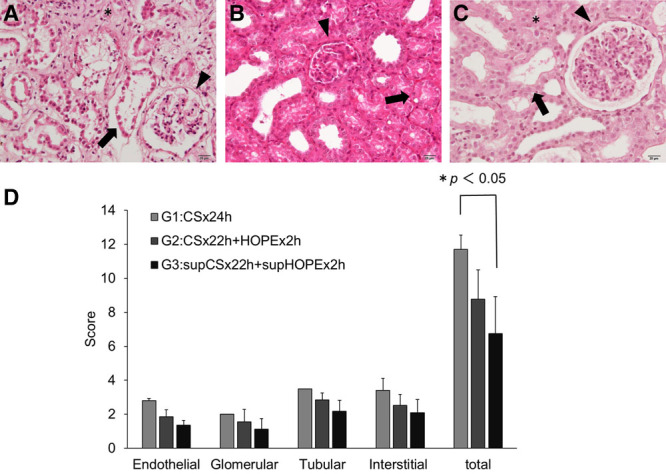
FIGURE 6.

Effects of quercetin and sucrose on the EGTI histology score in kidney tissue. Renal tissues were stained with hematoxylin-eosin (H&E), and pathological changes were evaluated using the EGTI scoring system under a light microscope at 400× magnification. A, H&E stain in G1 shows a glomerular tuft retraction (Glomerular score 2: ▾), complete necrosis in tubular cells (Tubular score 4: →), and inflammation and necrosis within the interstitium (Interstitial score 3: *). B, H&E stain in G2 shows a thickened Bowman’s capsule (Glomerular score 1: ▾) and thickened basal membrane of the tubular cells with loss of the brush border and the presence of cast formation (Tubular score 3: →). C, H&E stain in G3 shows the integrity of the basal membrane of the tubular cells with loss of the brush border in less than 25% of the tubular cells (Tubular score 1: →) and an intact glomerulus with thin-walled Bowman’s capsule (Glomerular score 0: ▾). There is no visible interstitium, signifying no damage/abnormality within the interstitial compartment (Interstitial score 0: *). In (D), the scoring graph shows data as the mean ± SD (G1: n = 2, G2: n = 4, G3: n = 4). Differences between each group were calculated using the Student’s t-test. G1:CS × 24h, G2:CS × 22h + HOPE × 2h, G3:supCS × 22h + supHOPE × 2h. CS, cold storage; EGTI, endothelial, glomerular, tubular, interstitial; HOPE, hypothermic oxygenated perfusion.
