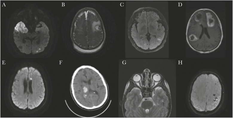
Figure 1.
Select neuroimaging findings. A, Patient 2: MRI of the brain without contrast, axial DWI image showing diffusion restriction in the right middle cerebral arteries and left posterior cerebral artery territories. B, Patient 5: MRI of the brain with and without contrast, axial T2 weighted image showing left frontal parenchymal hemorrhage with surrounding mass effect and nonspecific mild diffuse dural thickening. C, Patient 7: MRI of the brain with and without contrast, axial fluid attenuated recovery image showing left parietal juxtacortical white matter hyperintensity. D, Patient 17: MRI of the brain with and without contrast, axial T1 postcontrast image showing multifocal hypointensities with surrounding enhancement in the left frontal lobe, right temporoparietal lobe, and inferior right frontal lobe with surrounding vasogenic edema. E, Patient 23: MRI of the brain with and without contrast, axial DWI image showing subacute infarct in the left frontal centrum semiovale. F, Patient 25: CT of the head without contrast, axial image showing right thalamic intraparenchymal hemorrhage with associated mass effect on the right lateral ventricle. G, Patient 26: Axial MRI orbits with and without contrast: T2 weighted image showing mild kinking of the intraorbital optic nerves and mildly distended optic nerve sheaths bilaterally. H, Patient 27: Axial MRI of the brain without contrast: susceptibility weighted imaging showing hemorrhagic infarct of the left perirolandic region. Subacute subdural hematoma at the right frontoparietal lobe. Abbreviations: CT, computed tomography; DWI, diffusion weighted imaging; MRI, magnetic resonance imaging; SWI, susceptibility weighted imaging.

