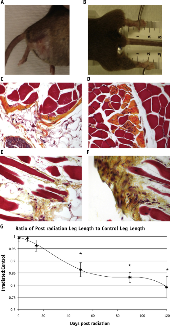
Fig. 1.
Fibrosis on gross and histologic examination. The irradiated right hind leg of a mouse has telangiectasias, alopecia, and fibrosis causing contracture at 90 days (A) and 120 days (B). Movat’s pentachrome stain shows elastin fibers in black, collagen fibers in yellow, proteoglycans in blue, muscle as red, and cell nuclei as purple. Unirradiated legs (C) and (D) do not show the increased collagen deposition and excess extracellular matrix muscular atrophy, and fibrosis as seen in the irradiated legs collected at 120 days (E) and (F). (G) *Significant leg shortening effects as shown by leg measurements at later timepoints: 50 (P=.027), 90 (P=.004), and 120 days (P=.013) after irradiation. A color version of this figure is available at www.redjournal.org.

