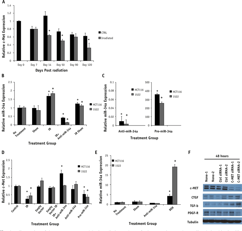
Fig. 4.
Expression of downstream target c-Met in radiation and transfection experiments show miR relationship. In silico analysis showed c-Met as a target of miR-34a and was therefore evaluated in vitro after irradiation (A). Time-points on days 14, 50, and 120 showed significantly decreased c-Met expression in comparison with time-matched controls (P.009, .057, and .059, respectively). Expression is normalized to the day 0 control tissue. HCT116 and 1522 cell lines (B-D) were transfected with empty vector (sham), anti-miR-34a, or pre-miR34a. Cells were also exposed to radiation (IR, 2 Gy: HCT116 and 10 Gy: 1522). Irradiation significantly increased miR-34a expression in both cell lines compared to control (CTRL) group (P=.024: HCT116, P=.011: 1522). This corresponded to a significant decrease in c-Met expression in the same cell lines (P=.017: HCT116, P=.032: 1522). Rescue transfection with anti-miR-34a in cells exposed to irradiation restored c-Met expression (B). This correlates with the restoration of c-Met expression (D) to a level comparable with control and in HCT116 cells greater than control (P=.047). Doxorubicin-treated cells caused a similar increase in miR-34a expression (P=.001: HCT116, P<.001: 1522) (E). Significance is noted by * for HCT116 and + for 1522 cells compared with respective no-treatment cells. Anti-c-Met siRNA transfection was completed to assess response of profibrotic molecules. Knockdown of c-Met in vitro in the 1522 cell line showed c-Met to be downregulated and an upregulation of TGF-β and CTGF by Western blotting (F).

