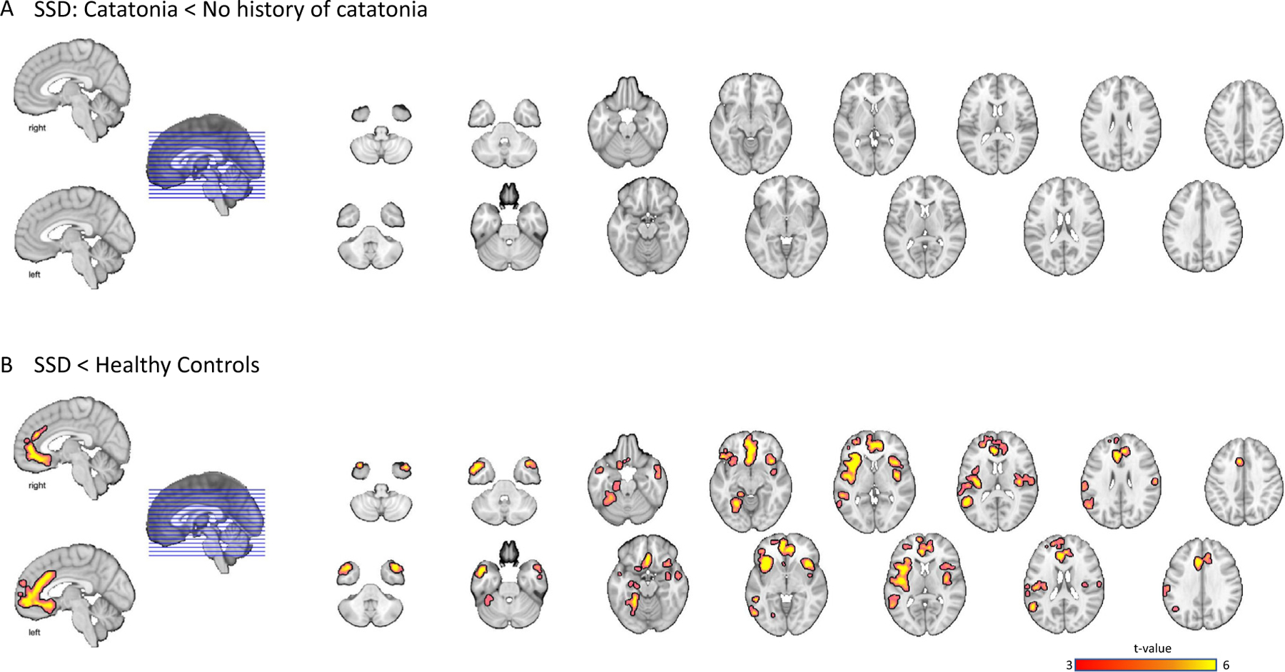
Fig. 3.

SSD with catatonia did not show grey matter differences compared to SSD without catatonia for both ROI and whole brain analysis (Panel A). A voxel-wise whole brain comparison showed that both patient groups combined had less grey matter volume in comparison to healthy control subjects in regions including anterior cingulate, medial frontal cortex, middle temporal gyrus and temporal pole, angular gyrus, and lingual gyrus (Panel B). Results were thresholded at cluster-level corrected p(FWE) = 0.05 for voxel-wise p(uncorrected) = 0.05. Images are presented in a neurological orientation.
