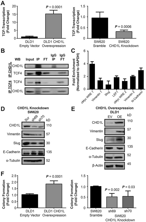
Figure 2: CHD1L mediates TCF-transcription in CRC.

(A) Overexpression of CHD1L in DLD1 cells shows an increase in TCF-transcription using the TOPflash TCF-transcription reporter assay (P < 0.0001), knockdown of CHD1L in SW620 cells using shRNA decreases TCF-transcription (P = 0.0006). Mean value of fold change from three independent experiments ± s.d. are shown. (B) Co-immunoprecipitation of CHD1L with TCF4 from SW620 cells, IP (immunoprecipitation), FT (flow-through). (C) Chromatin immunoprecipitation of CHD1L with WNT response element DNA promoter sites in SW620 cells. (D) Evaluation attenuated gene expression of EMT associated genes with SW620CHD1L-KD cells by Western blot. (E) Evaluation of induction of EMT gene expression in DLD1CHD1L-OE cells by Western blot. (F) Evaluation of CSC colony formation in DLD1CHD1L-OE, (P = 0.0001) and SW620CHD1L-KD (P = 0.002, 0.03). Mean values of fold change are from three independent experiments ± s.d. Representative colony images are shown in the supplemental information.
