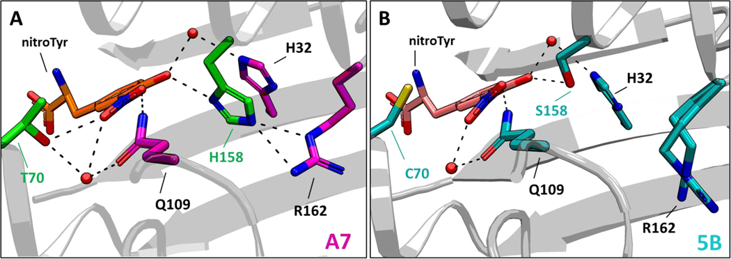
Figure 7.

Structural basis of nitroTyr recognition by the “A7” nitroTyr aaRS. (A) Crystal structure of the active site of the “A7” nitroTyr aaRS showing T70 and H158. In the “A7” active site, two additional hydrogen bonds from T70 and a bifurcated hydrogen bond from H158 to the guanidinium group of R162 are shown. (B) Active site configuration of the 2nd-generation “5B” nitroTyr aaR (PDB 4nda). R162 is shown in two equally populated conformations.
