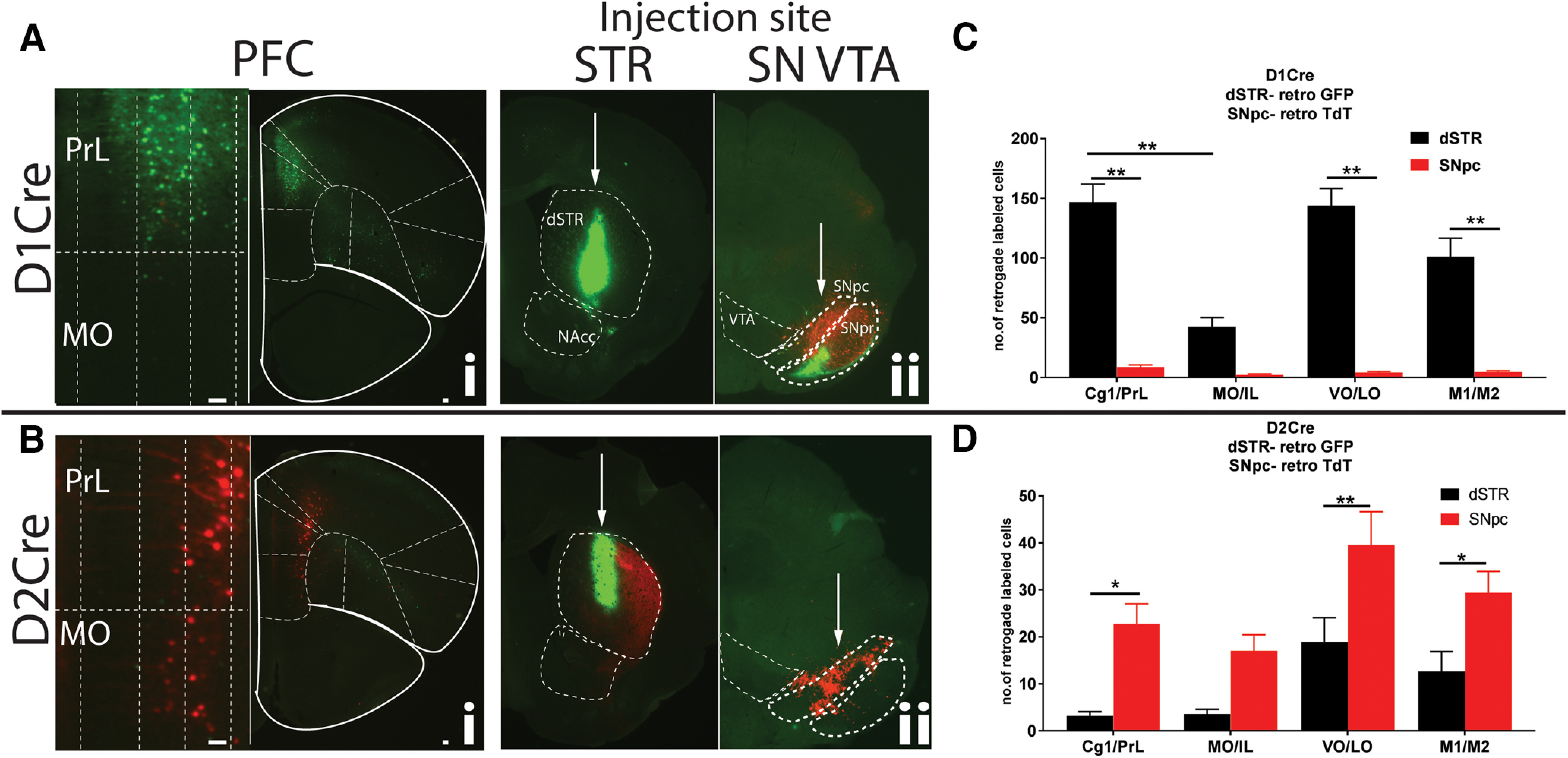
Figure 2.

Unique topographically organized subpopulations of D1R+ and D2R+ PFC neurons project to dSTR and SNpc. PFC sections from D1Cre (Ai) and D2Cre (Bi) mice showing cell bodies retrograde labeled with either GFP or TdTomato, respectively. Left panels, Zoomed insets of medial PrL and MO showing layer-specific localization of cell bodies. PrL, prelimbic; Cg1, anterior cingulate; MO, medial orbitofrontal; VO, ventral orbitofrontal; LO, lateral orbitofrontal; M1, primary motor; M2, secondary motor cortex. Aii, Bii, Injection sites are shown in striatal (STR) and midbrain SNpc/VTA (SN VTA) sections. D1Cre and D2Cre mice were injected with Cre-dependent retrograde AAV, AAVRG-CAG-Flex-TdTomato, and AAVRG-hSYN-DIO-GFP in dSTR (100 nl) and SNpc (40 nl). Representative images (n = 5). C, D, Quantification of number of cells. Data are represented as mean ± SEM; *p < 0.05, **p < 0.01, two-way ANOVA. Scale bar: 100 μm.
