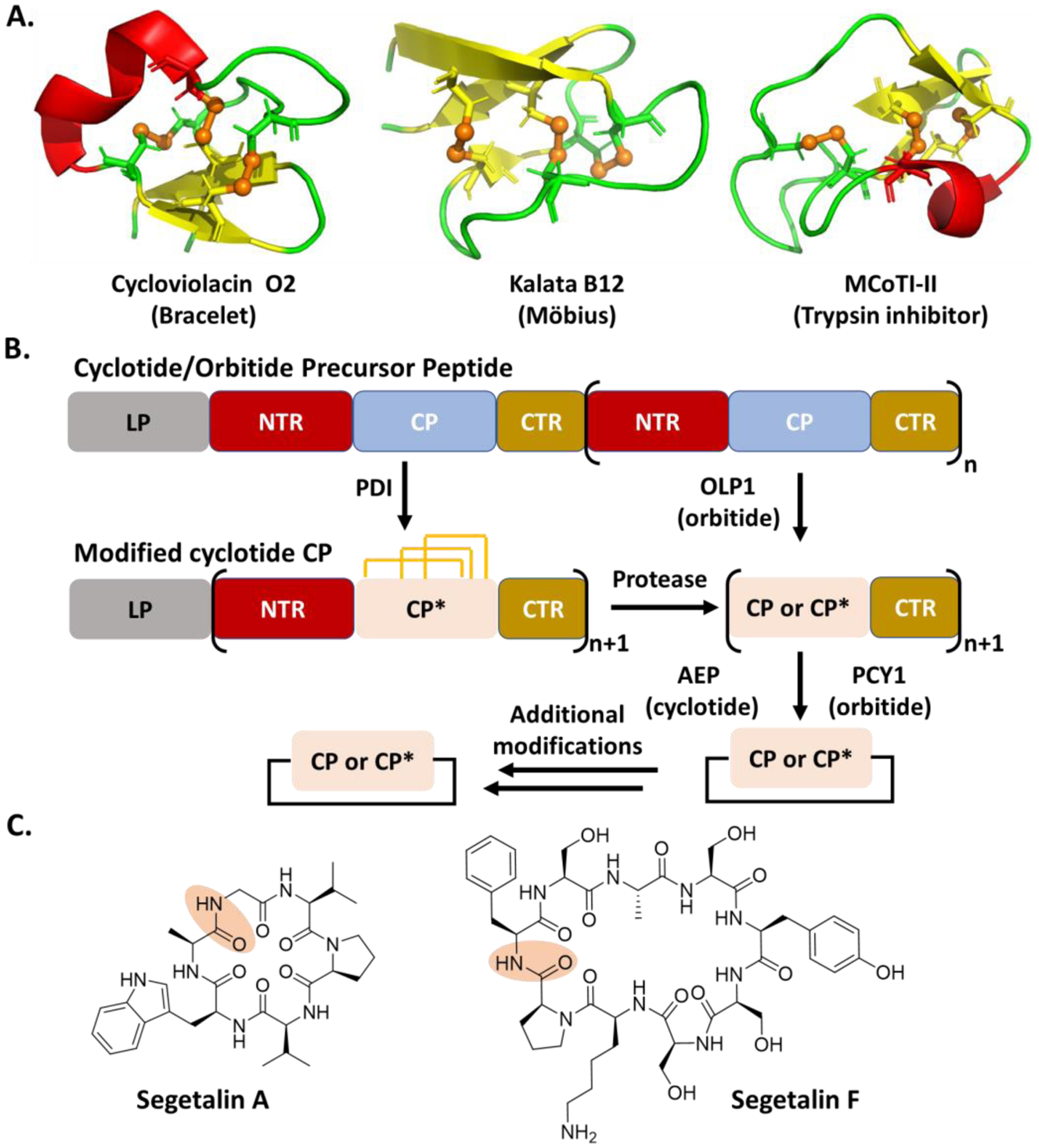
Fig. 6.

A. Representative cyclotides showing structural variations. The PDB IDs of Cycloviolacin O2, Kalata 12 and MCoTI-II are 2KCG, 2KVX, and 1HA9, respectively. The figures were created by Pymol. The disulfide bond is shown in brown and the brown sphere represents sulfur atom. B. General scheme of cyclotide and orbitide biosynthesis. The orange line represents a disulfide bond. C. Representative orbitide structures. The peptide bond for the cyclization is shadowed in orange.
