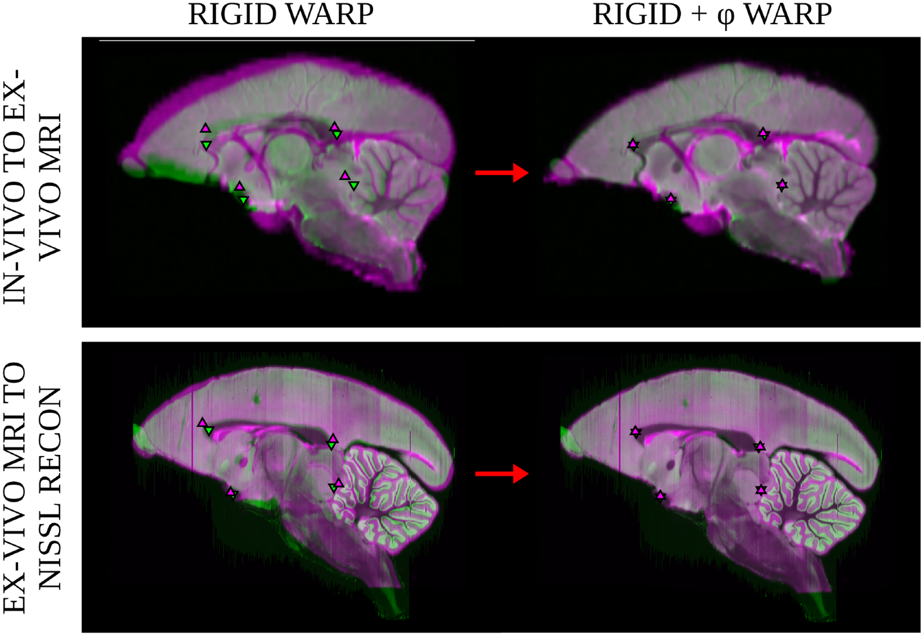
Figure 9: Image overlap after volume to volume registration.

Registration accuracy was measured to validate quantitative distortion measurements. The top row shows overlap of a subjectφs in-vivo MRI (magenta) mapped to the ex-vivo MRI (green) and the bottom row shows overlap of a subjectφs ex-vivo MRI (magenta) mapped to the nissl reconstruction (green). Landmarks located near the mid-sagittal plane from the landmark transfer analysis are overlayed on each image: posterior and anterior corpus callossum, anterior commissure, and the fastidium of the fourth ventricle.
