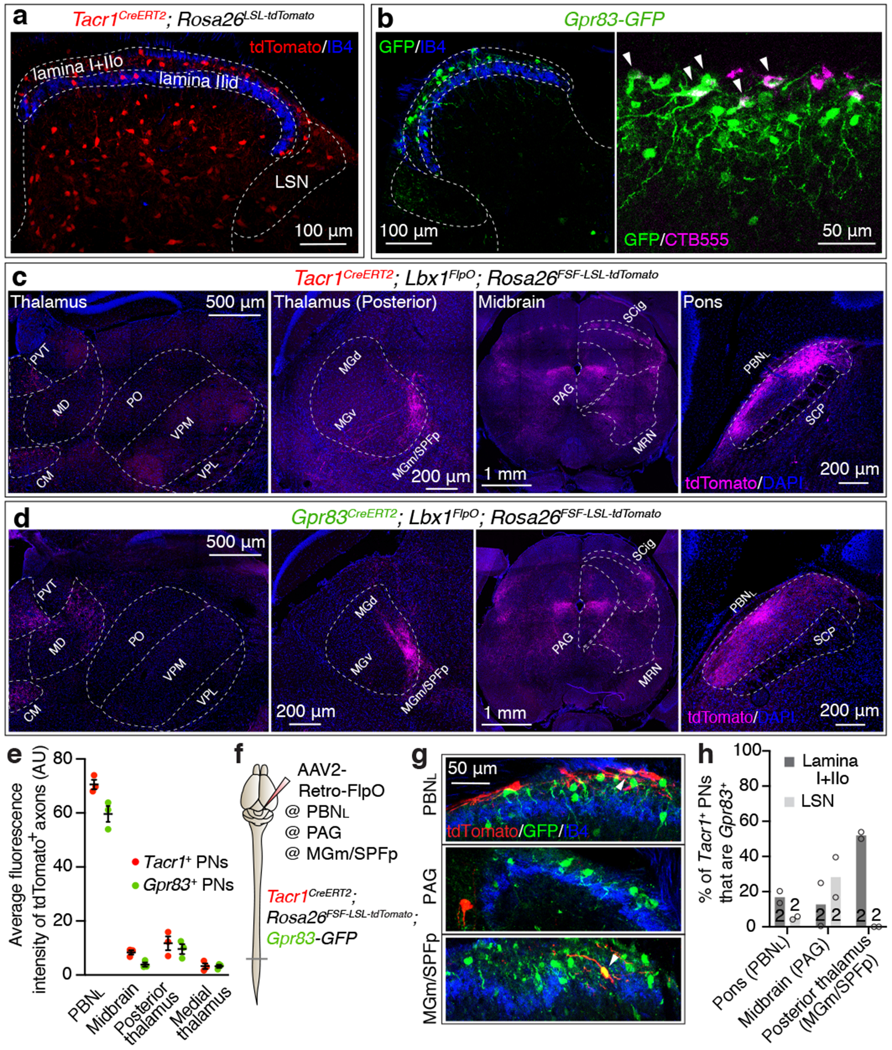
Figure 1. Tacr1- and Gpr83-expressing spinal PNs are largely distinct neuronal populations that innervate multiple distinct but overlapping brain regions.

a, Distribution of Tacr1+ neurons in the spinal cord dorsal horn. IIo, outer lamina II; IIid, inner dorsal lamina II; LSN, lateral spinal nucleus. b, Distribution of GFP-expressing Gpr83+ neurons in the spinal cord dorsal horn (left). A subset of SPB neurons labeled with CTB555 injected into the PBNL are GFP-positive (right). Arrow heads, double-positive neurons. c-d, Axonal projections of Tacr1+ or Gpr83+ spinal PNs. PVT, paraventricular nucleus; CM, central medial nucleus; MD, mediodorsal nucleus; PO, posterior complex; VPM, ventral posteromedial nucleus; MG(d)(v)(m), medial geniculate complex (dorsal)(ventral)(medial); SPFp, parvocellular subparafascicular nucleus; SCP, superior cerebellar peduncle. e, Quantification of the average fluorescence intensity of tdTomato-expressing Tacr1+ and Gpr83+ spinal PN axons in the major brain targets. n = 3 mice. Error bars, s.e.m. f, Schematic of virus injections for retrograde labeling of Tacr1+ spinal PNs. g, Distribution of tdTomato-expressing Tacr1+ spinal PNs and GFP-expressing Gpr83+ neurons in the spinal cord dorsal horn. Arrowheads, double-positive neurons. h, Quantification of co-expression of tdTomato and GFP. n = number of mice (indicated in the bar graphs).
