
Extended Data Figure 8. Anatomical analyses of axonal projections of anterolateral pathway PNs innervating the PBNL and the inferior olivary complex.
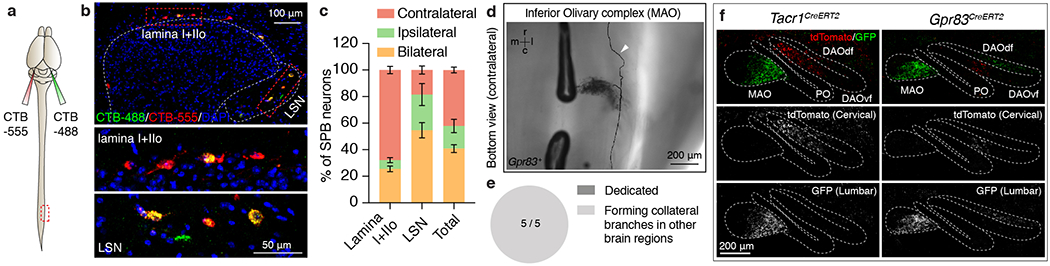
a, Schematic of dual-CTB injections into the PBNL. b, Distribution of CTB-labeled neurons in the spinal cord lamina I+IIo and the LSN. c, Quantification of % of SPB neurons that innervate the PBNL contralaterally, ipsilaterally, or bilaterally. n = 3 mice. Error bars, s.e.m. d, Bottom view of a single axon trace of sparsely labeled Gpr83+ spinal PN that innervate the inferior olivary complex. Arrowhead, an axon branch traveling up to the rostral brain. r, rostral; c, caudal, m, medial; l, lateral. e, Quantification of the number of inferior olivary complex-projecting spinal PNs that exhibit dedicated vs. collateral-forming axons. f, Synaptic terminals of Tacr1+ (h) or Gpr83+ (i) PNs representing hindlimb regions (GFP) and forelimb regions (tdTomato), are segregated in the inferior olivary complex. n = 3 mice each for Tacr1+ and Gpr83+ PNs.