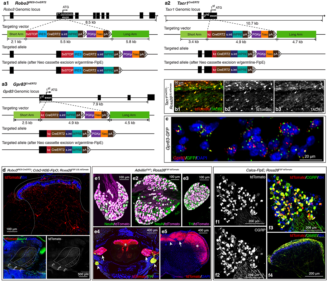
Extended Data Figure 1. Generation of CreERT2 mouse lines for genetic labeling of anterolateral pathway neurons and Flp mouse lines for labeling primary sensory neurons.

a1-a3, Gene targeting strategies used to generate the Robo3IRES-CreERT2 (a1), Tacr1CreERT2 (a2), and Gpr83CreERT2 (a3) mouse lines. a1, A 3X-STOP-IRES-CreERT2 cassette was introduced via homologous recombination into the first common coding exon that is shared by different splice variants of the Robo3 gene. a2, A CreERT2 cassette was introduced via homologous recombination into the Tacr1 gene, replacing the first coding ATG. a3, A CreERT2 cassette was introduced via homologous recombination into the Gpr83 gene, replacing the first coding ATG. IRES, internal ribosome entry site; s.int, synthetic intron; WPRE, Woodchuck Hepatitis Virus (WHP) Posttranscriptional Regulatory Element; pA, poly A; f, FRT site; kz, kozak sequence. b1-b3, A horizontal section of the lumbar spinal cord. 93.7 ± 2.6 % of tdTomato+ neurons were Tacr1+, while 96.6 ± 2.4 % of Tacr1+ neurons were tdTomato+. n = 3 mice. c, A transverse section of a Gpr83-GFP mouse. Green and red dots represent GFP and Gpr83 mRNA molecules, respectively, detected with gene-specific RNAscope probes. 96.0 ± 1.2% of GFP+ cells were Gpr83+, while 84.5 ± 5.0% of Gpr83+ cells were GFP+. n = 2 mice. d, Distribution of tdTomato-expressing Robo3+ neurons in the spinal cord dorsal horn (top) and their thalamic projections terminating in the VPL (bottom). n = 2 mice. e1-5, Characterization of the AdvillinFlpO mouse line. n = 4 mice. e1-e3, The AdvillinFlpO mouse line labels the majority of DRG neurons (99.0 ± 0.1% of NeuN+ neurons are tdTomato+) (e1), nodose ganglia neurons (80.8 ± 5.1% of NeuN+ neurons are tdTomato+) (e2), and sympathetic ganglia neurons (98.6 ± 0.3% of TH+ neurons are tdTomato+) (e3). e4, A transverse section of the vertebral column. tdTomato+ Advillin-expressing neurons and their axons are visualized in the spinal cord (asterisk), DRGs (arrows), and sympathetic ganglia (arrowheads). e5, A coronal section of the brainstem. tdTomato+ axons of Advillin-expressing neurons innervate the nucleus of the solitary tract (arrowhead), the dorsal column nuclei (arrow), and the trigeminal nucleus (asterisk). f1-4, Characterization of the Calca-FlpE mouse line. n = 2 mice. A cross section of the lumbar DRG (f1-f3) and a transverse section of the lumbar spinal cord (f4). f1-3, 91.9 ± 1.5 % of tdTomato+ neurons were CGRP+, while 92.3 ± 1.5 % of CGRP+ neurons were tdTomato+. f4, tdTomato-expressing axons of CGRP+ DRG neurons are CGRP immunoreactive in the spinal cord dorsal horn.
