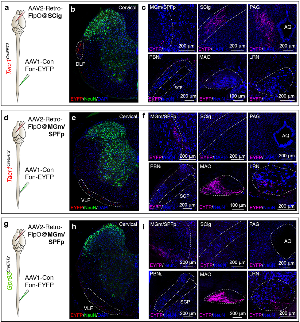
Extended Data Figure 3. Tacr1+ and Gpr83+ spinal PNs that innervate the posterior thalamus, midbrain, or the pons are distinct populations.

a, d, g, Schematics of lumbar spinal cord injections of AAV1-Con/Fon-EYFP viruses and brain injections of AAV2-retro-FlpO viruses into the SCig of Tacr1CreERT2 mice (a) (n = 3 mice), the MGm/SPFp of Tacr1CreERT2 (d) (n = 2 mice) or Gpr83CreERT2 mice (g) (n = 3 mice). b, e, h, Transverse sections of cervical spinal cords of Tacr1CreERT2 (b, e) or Gpr83CreERT2 mice (h). White dotted lines, tdTomato-expressing axons traveling through spinal cord white matter. DLF, dorsal lateral funiculus; VLF, ventral lateral funiculus. c, f, i, Coronal sections of target brain regions of Tacr1+ (c, f) or Gpr83+ (i) spinal PNs. AQ, cerebral aqueduct.
