
Fig. 2. The HACS1–PIRB interaction.
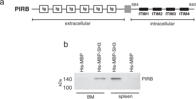
(a) Schematic representation of the PIRB receptor. The cytoplasmic region with four ITIM repeats spans approximately 190 residues. (b) Western detection of the HACS1-PIRB interaction. A pulldown was performed with immobilized HACS1 SH3 domain fragments fused to maltose-binding protein (MBP) and extracts of mouse bone marrow (BM) or spleen. Chemiluminescent detection of PIRB was achieved with a mouse anti-PIRB antibody.