Fig. 5.
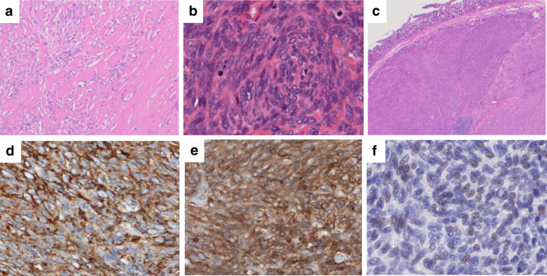
Histopathological findings of the resected specimen. Invasive growth of proliferated spindle-shaped cells in the pancreatic tumor. Hyalinized fibrosis is present at the periphery of the tumor (× 100) (a). The tumor showed high cellularity, increased mitotic figures (12/10 HPFs), and nuclear pleomorphism (increased N/C ratio) (× 200) (b). Tumor infiltration was observed on the duodenal side in contact with the main lesion (c). Immunohistochemically, the tumor cells were positive for CD34 (× 200) (d) and Bcl-2 (× 200) (e), and weakly positive for STAT6 (× 200) (f)

