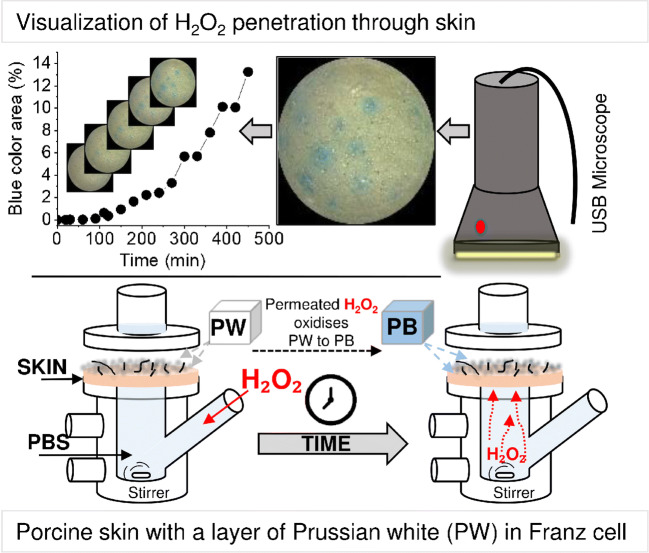
Scheme 1.
Schematic presentation of a skin membrane enclosed in a Franz cell. The skin was covered by PW microparticles. The lower chamber was filled with PBS containing H2O2 and 14 mM NaN3. With time, the H2O2 permeated the skin membrane and converted PW to PB. The development of blue colour was photographed using a USB microscope. The microscopy images revealed dominant H2O2 permeability pathways and the development rate of the blue area could be estimated

