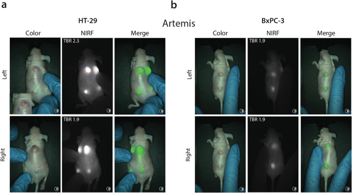
Fig. 5.
a Representative color, NIRF, and merged images of CH88.2-800CW binding specificity in a HT-29 tumor-bearing mouse model using the clinical Artemis NIR imaging system at 150-ms exposure. Regions of interest were selected in similar fashion to the Pearl as shown by the red and blue shapes, corresponding to the tumor and background area, respectively (only displayed in the left figure). To allow better visualization of the field of interest, the tumor-bearing skin was manually mobilized to the center of the camera’s optical field as is displayed by left and right back images. b Representative images of CH88.2-800CW binding specificity in a BxPC-3 tumor-bearing mouse model.

