
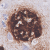
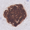
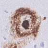
Table 2.
Staining summary per plaque-type
| Staining | The coarse-grained plaque | The cotton wool plaque | The classic cored plaque |
|---|---|---|---|
| Aβ (aa 8-17; 6F/3D) |
|
|
|
| H&E | Tissue distortion | Circumscript defined patches | Visible core |
| Congo red | ++; fibrillar amyloid throughout the plaque | −; not fibrillar | ++; fibrillar amyloid condensed into a core |
| pTau |
++; neuropil threads +−; dystrophic neurites |
++; neuropil threads | +−; dystrophic neurites |
| APP | +; dystrophic neurites | +− | +; dystrophic neurites |
| ApoE | ++; throughout the plaque | ++; throughout the plaque | ++; in the core; +−; in the corona |
| PrPC | +; throughout the plaque | +; throughout the plaque | ++; in the core; +−; in the corona |
| Aβ40 | ++; throughout the plaque as fibrillary or tubular structures, 61% as shell surrounding the lesser Aβ42 | ++; homogenous throughout the plaque | ++; in the core; +−; in the corona |
| Aβ40 | +−; in the plaque center, sometimes co-localizing with Aβ40 | +; outer ring | ++; in both the core and the corona |
| AβN3pE | ++ | ++ | ++ |
| pSer8Aβ | ++ | ++; intense stained plaque with diffuse halo | ++ |
| C4b | ++; throughout the plaque | +; outer ring | +; in the core; +−; in the corona |
| CD68 | ++; within Aβ-devoid pores | +−; occasionally 1 cell body within the plaque | +; in-between the core and the corona |
| MHC-II | ++; within Aβ-devoid pores | +−; occasionally 1 cell body within the plaque | +; in-between the core and the corona |
| GFAP | +; cell bodies are often found within the plaque | +; disrupted processes mostly staining the outer plaque edges | + |
| Norrin | ++; fibril-like throughout the plaque | ++; homogenous throughout the plaque | − |
| Laminin | +; small punctate dots throughout the plaque | +; small punctate dots throughout the plaque | +; small punctate dots surrounding the core |
| Collagen IV | − | − | +−; small punctate dots surrounding the core |
(Immuno)histochemical staining summary per plaque-type is given and indicated as follows: −, no staining; +−, some positive staining; +, positive staining; ++, prominent positive staining. Supplementary Material 1, Table S1, Online Resource, for antibody and staining details