
Figure 1.
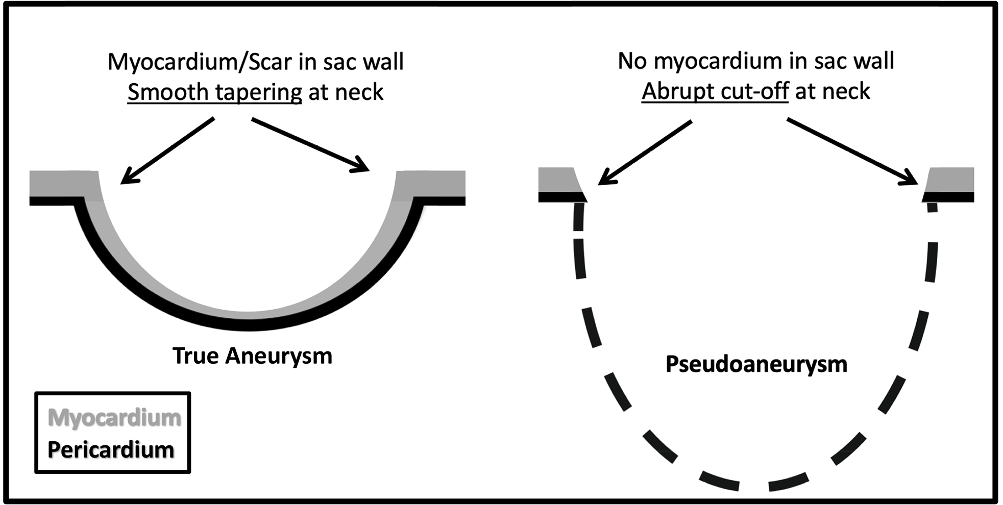
Diagram of the differences between a true left ventricular aneurysm (LVA) and pseudoaneurysm (LV PSA), which illustrate the concept of the myocardial cut-off sign. There is myocardium/scar tissue in the wall of LVA such that there is a more gradual tapering of sac wall thickness. LV PSA is a contained rupture and there is no myocardium in the sac wall. An abrupt “cut-off” of myocardium at the sac neck is present in LV PSA.