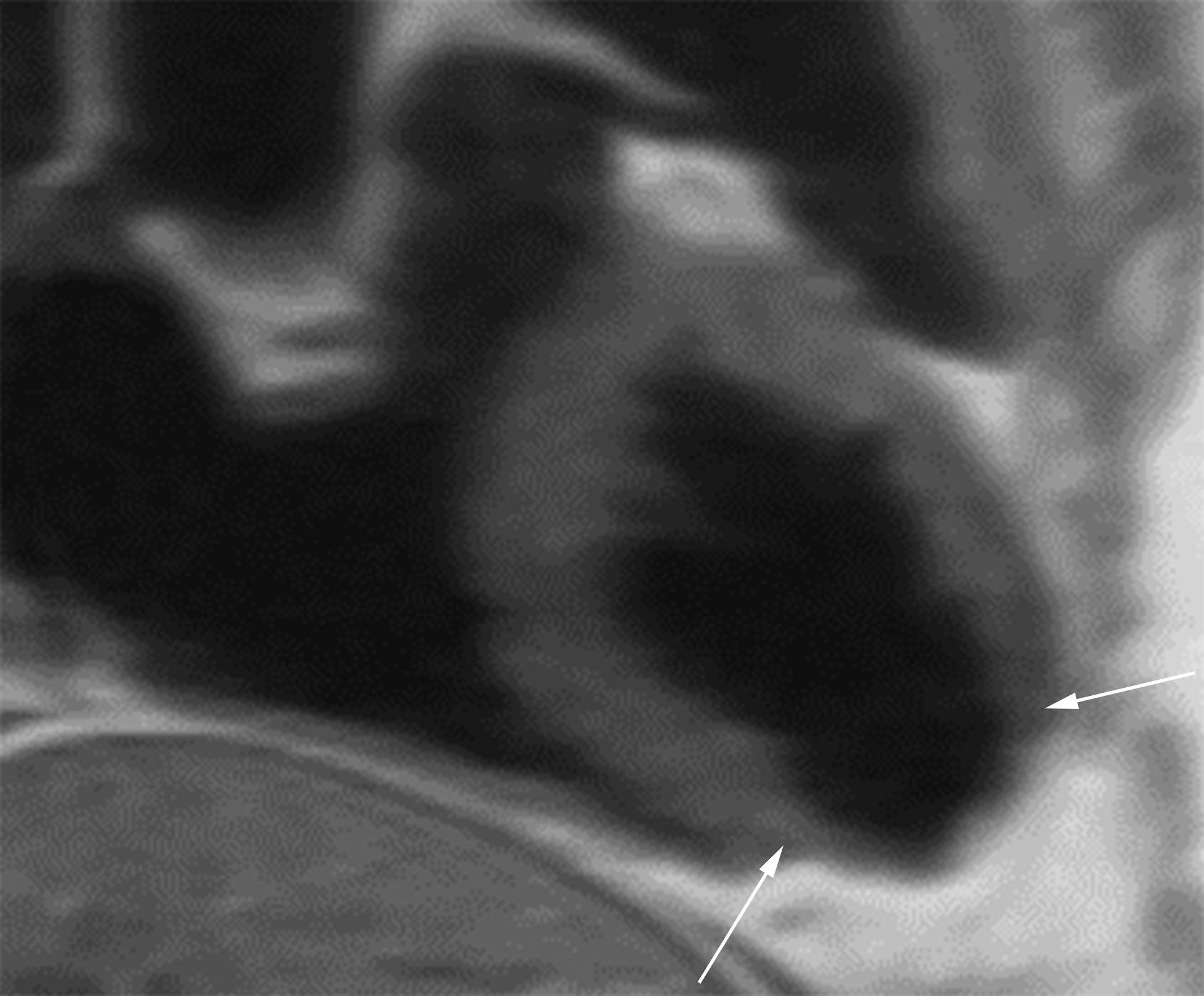
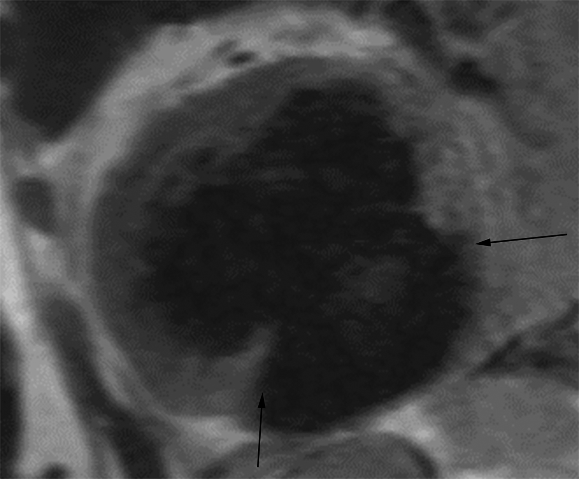
Figure 3.


Oblique two-chamber long axis black-blood cardiac MR image (CMRI) (a) from a patient with an apical true LVA demonstrates relatively gradual tapering of the ventricular wall at the sac neck (white arrows). Short-axis black-blood CMRI (b) from a patient with an inferior wall LV PSA demonstrates abrupt loss of myocardial thickness at the sac neck (black arrows).
