
Figure 6.
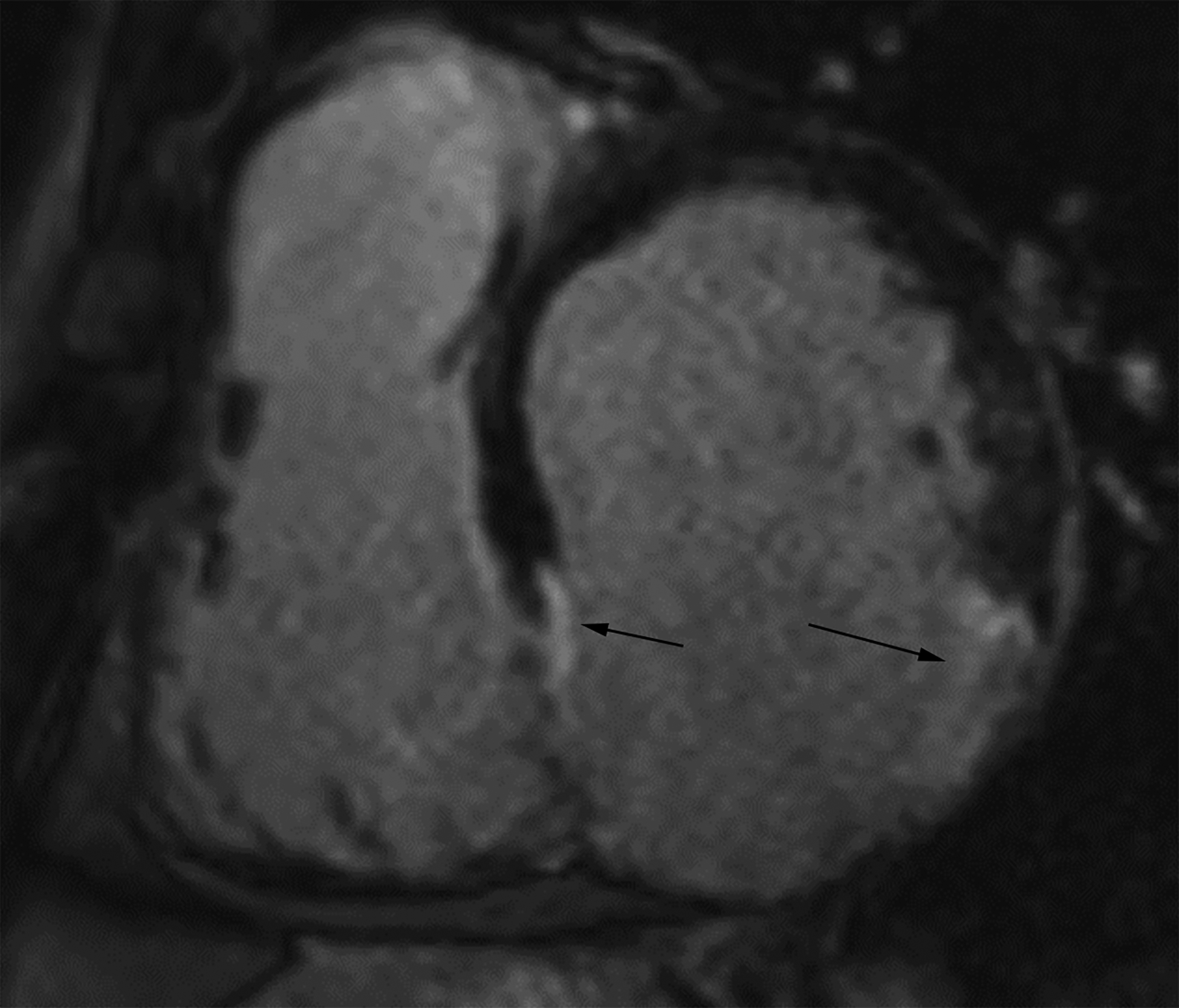
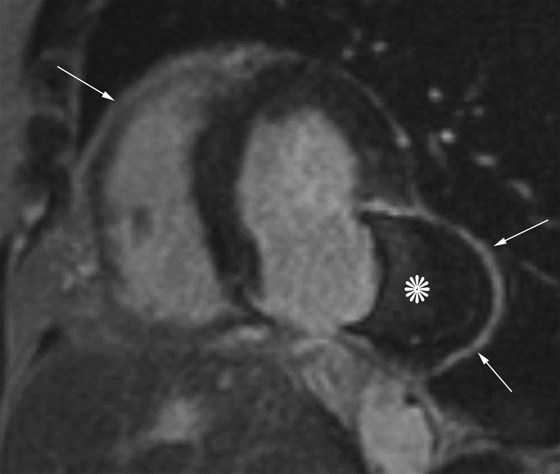
Short axis delayed enhanced CMRI (a) from a patient with an inferior wall true LVA (same patient as Figure 4a) demonstrates enhancing myocardial scar tissue within the sac wall (black arrows). Short axis delayed enhanced CMRI (b) from a patient with an inferolateral LV PSA demonstrates prominent pericardial enhancement (white arrows). Also note the thrombus filling much of the sac (*).