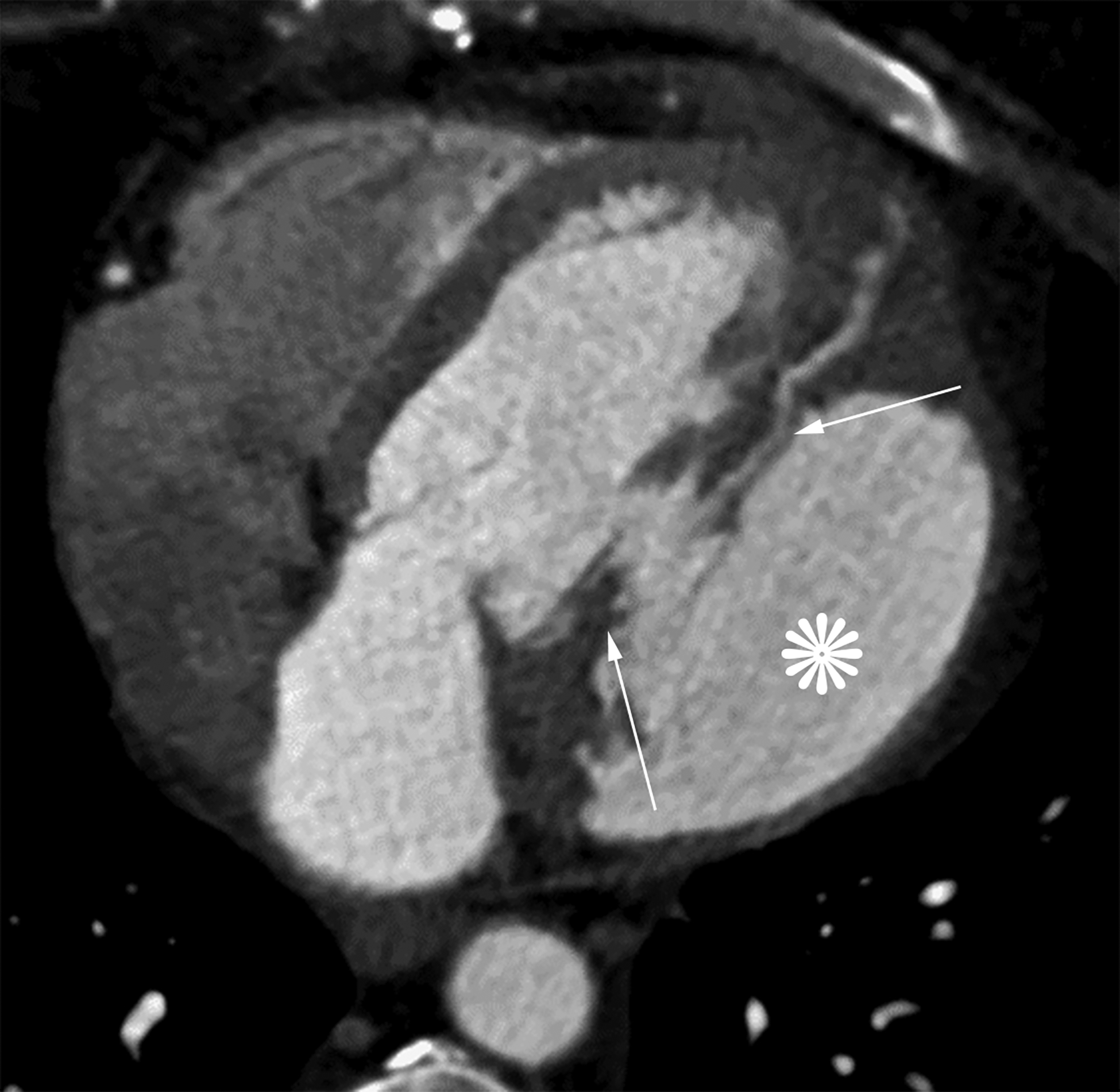
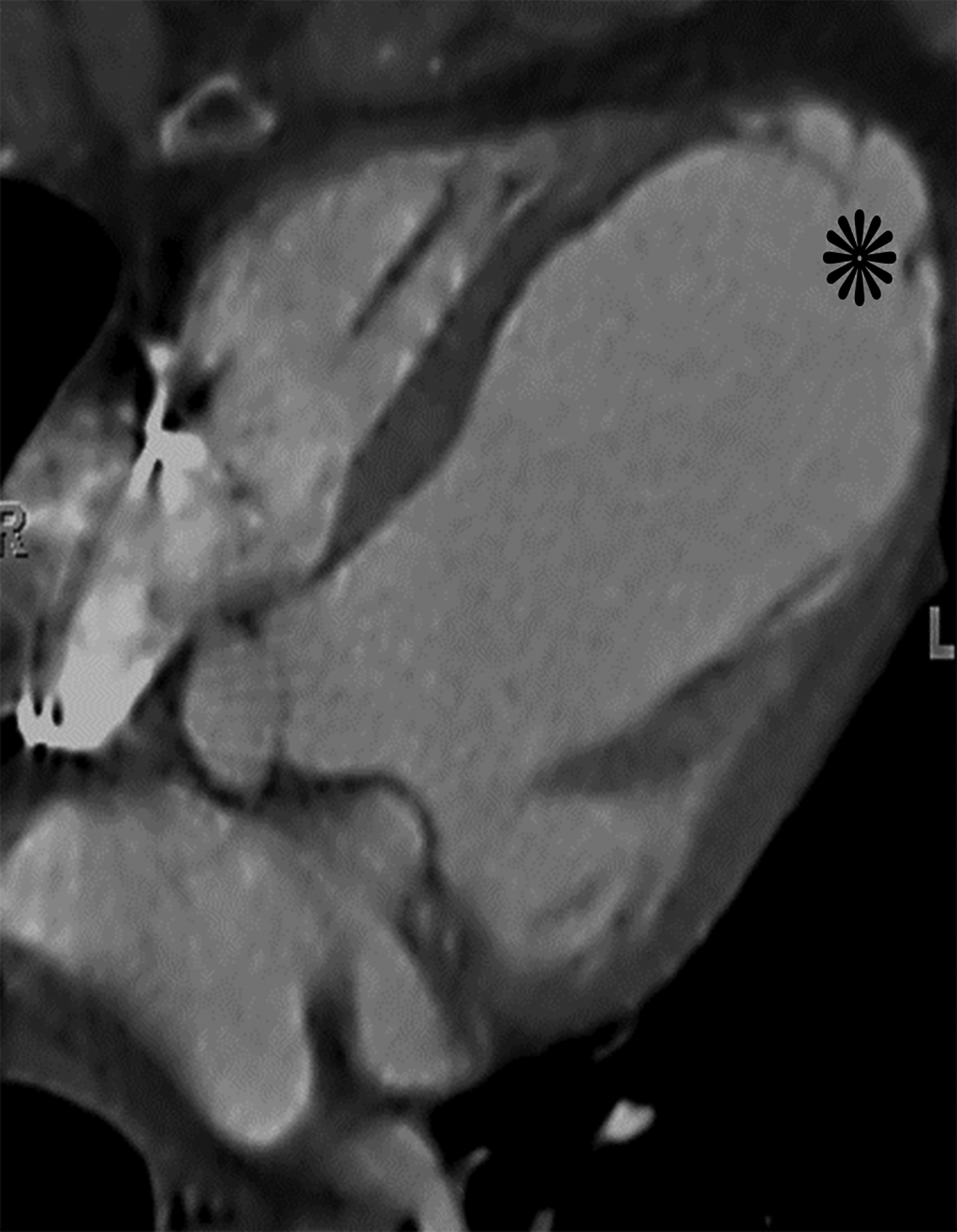
Figure 8.


Axial oblique contrast-enhanced CT (a) from a patient with a prior lateral wall LV PSA status post patch repair. The patch (arrow) had dehisced resulting in a recurrent LV PSA (white *). Note the narrow neck relative to the sac size. Axial oblique contrast-enhanced CT (b) from a patient with a large left anterior descending infarct with resultant apical true LVA (black *). Note the relatively wide neck. Also notice the smooth tapering of myocardium near the sac neck.
