4.
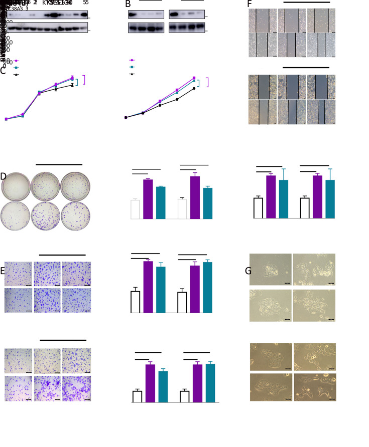
Loss of SLC38A3 promotes cell proliferation, migration and invasion of ESCC. (A) Western blotting analysis of SLC38A3 in nine ESCC cell lines and two immortalized normal esophageal cell lines NE2 and NE3; (B) Western blotting analysis of SLC38A3 expression after introducing small interfering RNA (siRNA) of SLC38A3 in KYSE410 and KYSE510. Cell lysates were collected 48 h after transfection; (C) Cell proliferation assays were performed after introducing siRNA of SLC38A3 in KYSE410 and KYSE510, and cell viability was quantified by MTS assay. Data represent
 , n=3 independent experiments. Unpaired t test was performed to calculate P values; (D) Colony formation assays with siCtrl, siSLC38A3_1, siSLC38A3_3 transfected cells for 10 d. Data represent
 , n=3 independent experiments. Unpaired t test was performed to calculate P values; (E) Transwell-based migration and invasion assay with siCtrl, siSLC38A3_1, siSLC38A3_3 transfected cells for 12 h. Scale bars: 50 µm. Data represent
 , n=3 independent experiments. Unpaired t test was performed to calculate P values; (F) Wound healing assay of siCtrl, siSLC38A3_1, siSLC38A3_3 transfected cells for 24 h. Representative images were taken with phase contrast microscopy. Scale bars: 50 µm; (G) Representative images of siCtrl, siSLC38A3_1 transfected cells with or without TGF-β (10 ng/mL) for 48 h. Scale bars: 50 µm. **, P<0.01; ***, P<0.001; ****, P<0.0001 for three independent experiments analyzed by Student’st test. SLC38s, solute carrier family 38; ESCC, esophageal squamous cell carcinoma; TGF-β, transforming growth factor beta.

