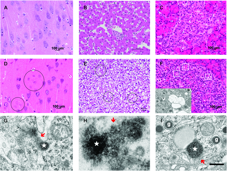
FIGURE 6.
Cell degeneration/death and lysosomal rupture in hippocampal CA1 neurons, hepatocytes, and pancreatic β-cells. A, B, C: hematoxylin-eosin staining of the hippocampus (A), liver (B), and pancreas (C) of a control monkey without hydroxynonenal injections. D, E, F: in monkeys after sublethal doses of hydroxynonenal injections, neuronal loss was observed in the CA1 (D, circles), whereas ballooning and cell death (E, circles) of hepatocytes, and microcystic degenerations of the Langerhans cells (F, white rectangles) were seen. The cells with microcystic degenerations were confirmed by electron microscopy to be degenerating β-cells, because of the presence of insulin granules with halo (F, inset). G, H, I: the degenerating CA1 neurons (G) and hepatocytes (H) showed lysosomal permeabilization/rupture with spillage of the lysosomal content (red arrows) from the lysosome (star), whereas β-cells (I) with insulin granules (g) showed permeabilization (red arrow) of lysosome (star) in the process of fusing with an autophagosome. Bar = 400 nm (G, H, I). Note that such lysosomal disintegrity of the hydroxynonenal monkeys was quite similar to that of a human patient with Alzheimer's disease as shown in Figure 3C. (Adapted with permission from reference 14.)

