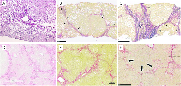
FIGURE 1.
Histopathology of human NASH and models with advanced fibrosis following a Western diet. (A) Perisinusoidal fibrosis (F1) located around a central vein in a patient with NASH (Sirius Red Hemalun stain, ×10 magnification) Modified from reference 14 with permission. (B) Bridging fibrosis (F3) and (C) cirrhosis (F4) with thick and extensive bridging in patients with NASH. Arrows denote fibrous bridges and arrowheads indicate perisinusoidal fibrosis, P: portal tract, V: hepatic vein (Elastica van Gieson stain; scale bar, 250 μm). Modified from reference 15 with permission from Wiley. (D) Bridging fibrosis in the DIAMOND mouse (Sirius Red stain, ×10 magnification). Modified from reference 16 with permission. (E) Bridging fibrosis in the guinea pig NASH model also showing the prototypical perisinusoidal (chicken-wire) pattern (Picro Sirius Red stain and Weigert's hematoxylin; scale bar, 200 μm). (F) Bridging fibrosis (arrows) with perisinusoidal collagen deposition in hamsters, inset is part of the original figure, but is not used in this reproduction (Sirius Red stain, ×10 magnification). Modified from reference 17 with permission. Permissions to reuse have been obtained for all figures and the documentation is provided in the Supplemental Material. DIAMOND, diet-induced animal model of nonalcoholic fatty liver disease; NASH, nonalcoholic steatohepatitis.

