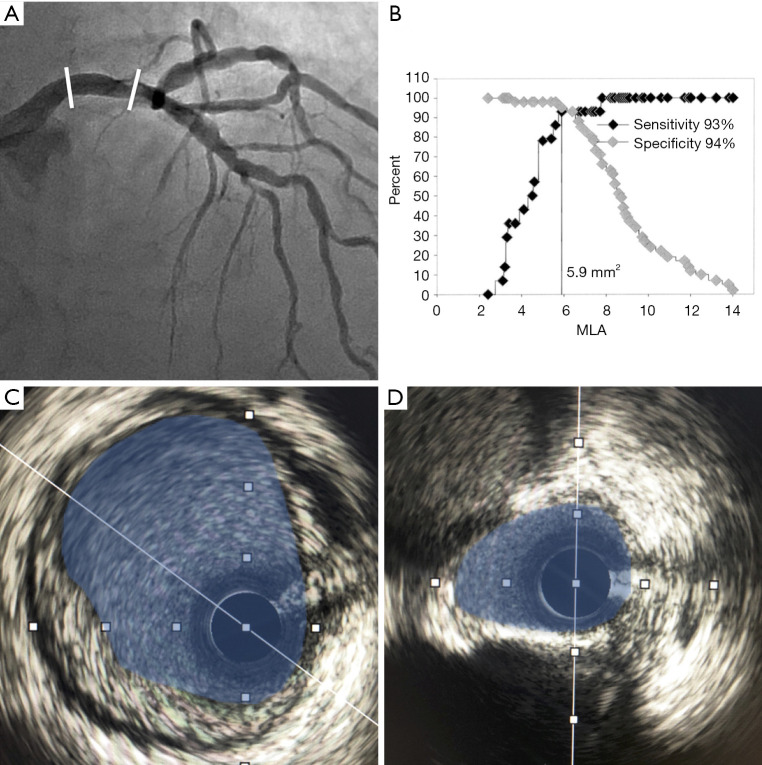
Figure 1.
Intravascular ultrasound assessment of an intermediate left main coronary artery stenosis. Eccentric and calcified distal left main coronary artery (LMCA) lesion (A). Representative cross-sectional intravascular ultrasound (IVUS) images of the proximal reference segment with highlighted minimal luminal area (MLA) (highlighted blue) (C) and target lesion MLA (blue) highlighting eccentricity and superficial calcification (D). The calculated MLA (shaded blue) was 5.1 mm2, which is below the safe deferral threshold of 6 mm2 (B). Panel B reproduced from Jasti et al. Circulation, 2004 (32).

