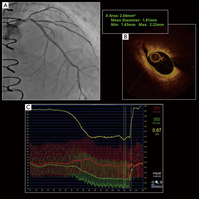
Figure 4.
Optical coherence tomography and fractional flow reserve. Intermediate lesion of the distal left main coronary artery (A), which extends into the proximal left anterior descending (LAD). Initial optical coherence tomography images (B) demonstrate a minimal luminal area of 2.64 mm2, with evidence of a small intimal dissection (at 11 o’clock). Fractional flow reserve (FFR) assessment of the vessel (C) demonstrates that the lesion is highly haemodynamically significant (FFR 0.67).

