FIG 5.
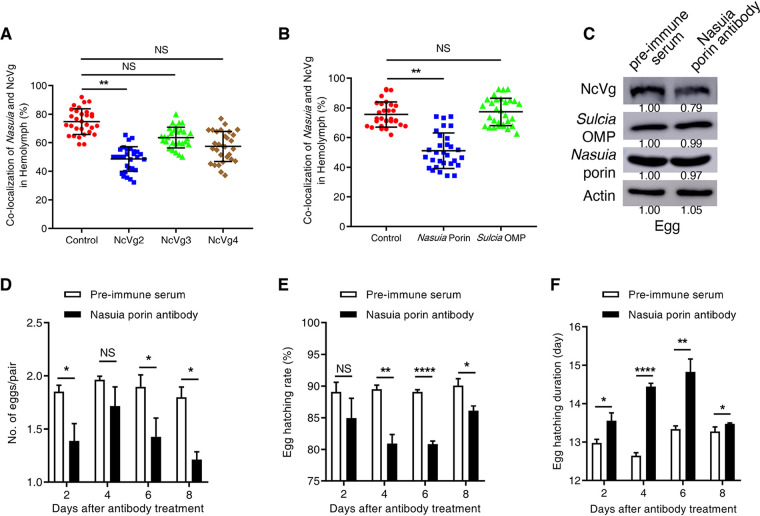
NcVg transport into oocytes is mediated by Nasuia. (A) Confocal microscopy showed the reduced colocalization of Nasuia with NcVg in the hemolymph of female adults after the treatment with NcVg2 antibody rather than with NcVg3 or NcVg4 antibodies. (B) Confocal microscopy showed the reduced colocalization of Nasuia with NcVg in the hemolymph of female adults after the treatment with Nasuia porin antibody rather than with Sulcia OMP antibody. The hemolymph smears of insects treated with NcVg antibodies (A) or symbiont antibodies (B) were stained with Nasuia-cy3 and NcVg-FITC and observed by confocal microscopy. The number of fluorescent spots that were positive for both cy3 and FITC was calculated and represented colocalization of NcVg and Nasuia. (C) Western blot assay showed the constant levels of Nasuia porin and Sulcia OMP in porin antibody-treated eggs laid by antibody-treated adult females but decreased NcVg levels in treated eggs. The protein accumulation levels of NcVg, Nasuia porin, and Sulcia OMP in the eggs of N. cincticep that received preimmune serum were taken to be 1.00. (D) The numbers of eggs laid by adult females treated with porin antibody or preimmune serum were comparable. (E and F) Nasuia porin antibody treatment significantly reduced egg hatching rate (E) and prolonged the egg hatching duration (F). Data in panels A, B, and D to F are presented as means ± SD of three independent experiments. The significance of any differences was tested using an independent t test. *, P < 0.05; **, P < 0.01; ****, P < 0.0001; NS, not significant.

