
FIG 6.
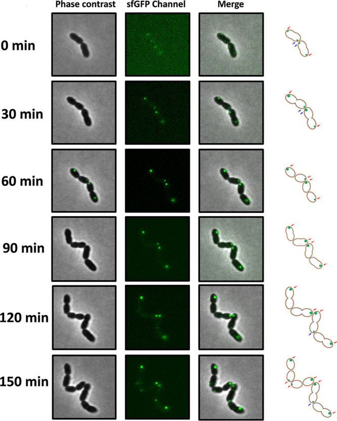
The nisin modification complex represented by NisB-sfGFP was primarily localized to the old pole. Cells from the culture of the strain NZ9000/pTLR3-nisA-nisBsfgfp-nisTC were transferred to a microscope slide with an agarose patch containing growth medium with 5 ng/ml nisin Z as an inducer. Images were captured at 15-min intervals using time-lapse microscopy. The red arrows indicate the appearance of old fluorescent poles. The blue arrows show the appearance of new fluorescent poles.