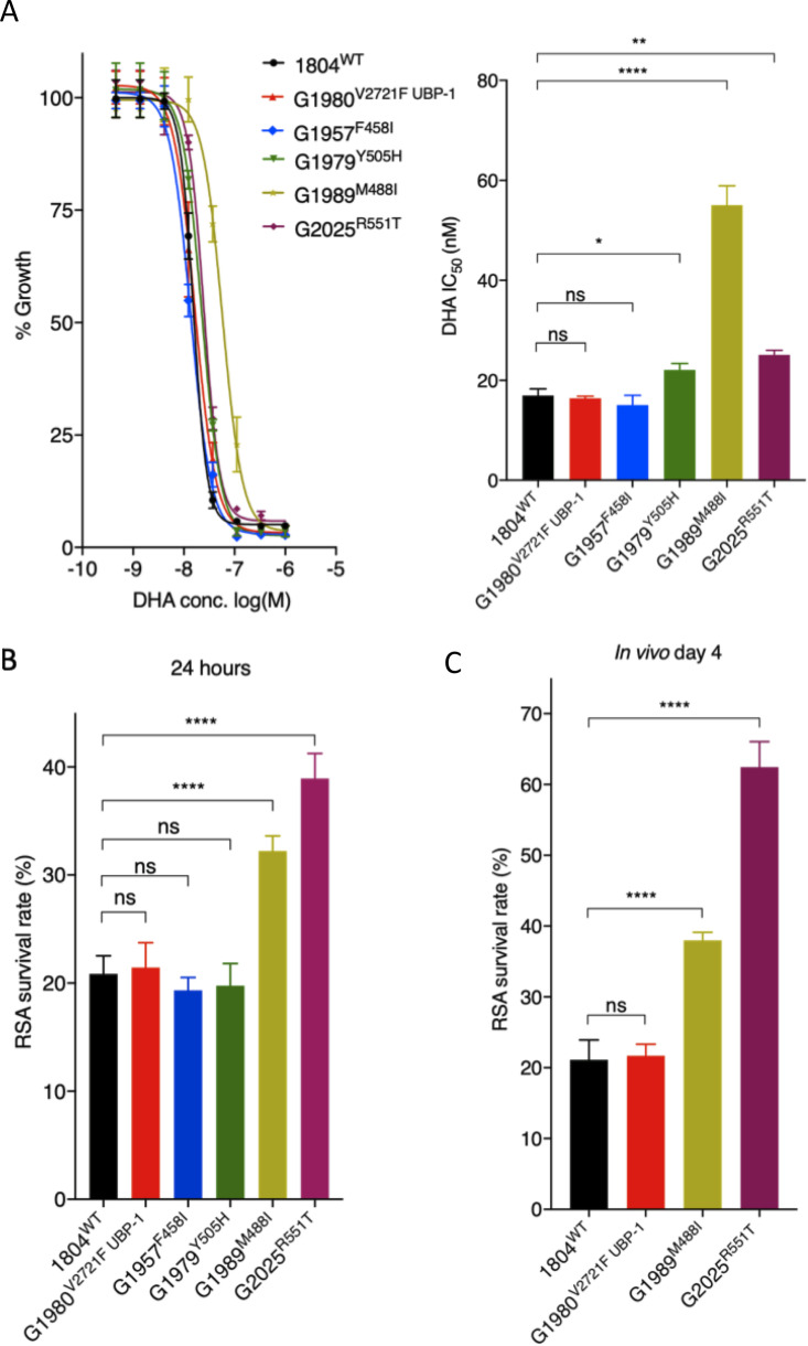
FIG 2.
In vitro and ex vivo susceptibility of P. berghei K13 mutants to DHA. (A) DHA dose-response curves and IC50 values for P. berghei K13 mutant lines compared to those of the wild-type 1804WT and the UBP-1 G1980V2721F mutant lines. (B) Survival of P. berghei K13 mutant lines in the P. berghei RSA. Results show the percentages of synchronized early ring-stage parasites (1.5-h postinvasion) that survived a 3 h exposure to 700 nM DHA relative to DMSO-treated parasites. Survival was quantified 24 h posttreatment by flow cytometry analysis based on Hoechst 33258 DNA staining and mCherry expression. (C) In vivo RSA survival for two K13 mutant lines (G1989M488I and G2025R551T) compared to that of the wild-type (1804WT) and UBP-1 mutant (G1980V2721F) controls. After in vitro exposure to DHA or DMSO as described above, parasites were i.v. injected back into mice as described in Materials and Methods. Parasitemia was quantified by flow cytometry analysis of mCherry expression on day 4 after i.v. injection, from which percentage survival rates were calculated. Error bars show standard deviations calculated from three biological repeats. Statistical significance (compared to the 1804WT line) was calculated using one-way analysis of variance (ANOVA) alongside the Dunnett’s multiple-comparison test. ns, not significant; *, P < 0.05; **, P < 0.01; ****, P < 0.0001.

