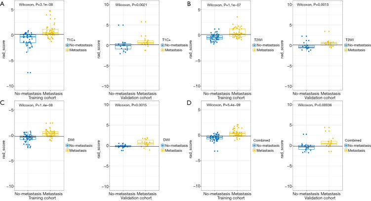
Figure 3.
Comparison of the Rad-scores from T2WI, DWI, T1C+ and combined model between LNM and non-LNM group were shown in A-D. In all the four models, the Rad-scores of LNM patients were significantly higher than those of non-LNM patients in both training and validation cohort. T2WI, T2 weighted imaging; DWI, diffusion weighted imaging; T1C+, T1 weighted multiphase contrast enhancement imaging; LNM, lymph node metastasis.

